Retinitis Pigmentosa 63

For a general phenotypic description and a discussion of genetic heterogeneity of retinitis pigmentosa, see 268000.
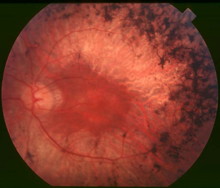
Clinical FeaturesKannabiran et al. (2012) described a large Indian family in which 14 of 34 individuals studied had retinitis pigmentosa. Age at presentation ranged from 16 to 65 years, and in most cases the initial symptoms consisted of night blindness associated with blurred vision. Fundus examination revealed a range of features, including degeneration of the retinal pigment epithelium (RPE) to varying extents, arterial attenuation, disc pallor, and pigment migration. Electroretinography (ERG) showed diminished or extinguished responses. The central retina was relatively less involved in most of the affected individuals, as suggested by good visual acuities and fundus appearance, with peripheral visual field loss and severe peripheral retinal involvement.
MappingKannabiran et al. (2012) genotyped 34 members of a large 4-generation Indian family segregating autosomal dominant retinitis pigmentosa (RP) for microsatellite markers located in proximity to known loci for autosomal dominant retinal dystrophy phenotypes, and obtained a maximum lod score of 2.9 (theta = 0) for marker D6S262 on chromosome 6q23. Fine mapping with additional markers yielded 2 markers, D6S457 and D6S1656, with maximum lod scores of 3.8 (theta = 0). Haplotype analysis in this family indicated a disease-cosegregating region of 28 cM (34 Mb). Kannabiran et al. (2012) stated that the mapped interval contained more than 100 genes, among which there were no known RP genes.