Hypertrophic Osteoarthropathy, Primary, Autosomal Recessive, 1

A number sign (#) is used with this entry because of evidence that autosomal recessive primary hypertrophic osteoathropathy-1 (PHOAR1) is caused by homozygous mutation in the HPGD gene (601688) on chromosome 4q34.
Isolated digital clubbing (119900) as well as cranioosteoarthropathy can also be caused by homozygous mutation in the HPGD gene.
DescriptionPrimary hypertrophic osteoarthropathy is a familial disorder characterized by digital clubbing and osteoarthropathy, with variable features of pachydermia, delayed closure of the fontanels, and congenital heart disease. Secondary hypertrophic osteoarthropathy, or pulmonary hypertrophic osteoarthropathy, is a different disorder characterized by digital clubbing secondary to acquired diseases, most commonly intrathoracic neoplasm (Uppal et al., 2008).
Touraine et al. (1935) recognized pachydermoperiostosis as a familial disorder with 3 clinical presentations or forms: a complete form characterized by periostosis and pachydermia; an incomplete form with bone changes but without pachydermia; and a 'forme fruste' with pachydermia and minimal skeletal changes.
Genetic Heterogeneity
PHOAR2 (614441) is caused by mutation in the SLCO2A1 gene (601460) on chromosome 3q22.1-q22.2.
Families with an autosomal dominant form of primary hypertrophic osteoarthropathy have also been reported (PHOAD; 167100).
Clinical FeaturesFriedreich (1868) first described pachydermoperiostosis in 2 brothers, Wilhelm and Carl Hagner, both of whom had had onset in their teens. They had 4 unaffected sibs.
Vogl and Goldfischer (1962) described the clinical features of pachydermoperiostosis as including clubbing of the fingers, thickening of the skin and periosteum of the distal part of the extremities, thickening and seborrhea of the skin of the face and forehead, and hyperhidrosis.
Hedayati et al. (1980) reported a 45-year-old black woman with classic features of pachydermoperiostosis, including clubbing of the digits, periosteal new bone formation, and hypertrophy of the skin associated with severe facial acne. In addition, she showed excessive resorption of distal phalanges of the hands and feet, which had not previously been reported in pachydermoperiostosis. Hedayati et al. (1980) postulated that reduced peripheral blood flow may have resulted in bone resorption.
Matucci-Cerinic et al. (1989) described a 19-year-old man with PDP whose symptoms began at age 17. He had digital clubbing, pachydermia, and generalized hyperhidrosis. The palms and soles showed profuse and continuous sweating. Radiographs showed periostosis of the diaphyses of the long bones, but acroosteolysis was not evident. Studies of blood flow in the fingers were normal, but there was evidence of irregularly shaped capillaries and new capillary formation. Skin biopsies from affected areas showed diffuse endothelial hyperplasia, hyalinosis, and sclerosis with abnormalities of collagen fibers. The father had mild finger clubbing without other symptoms or signs of the disorder.
Sayli et al. (1993) described PDP in 3 males out of 7 sibs from a village in mid-Anatolia. The parents were second cousins, suggesting autosomal recessive inheritance. The 3 affected males were aged 14, 12, and 10 years; ulcers had been present from the age of 3 or 4 years. Below-the-knee amputation was performed in the oldest of the affected brothers. The disorder was thought to differ from the autosomal dominant form by the presence of growth retardation, early ulcers, and acrolysis of the distal parts of the extremities with secondary contractures.
Singh and Menon (1995) described a 13-year-old boy with progressive enlargement of the joints and distal extremities, clubbing, coarse facial features, and hyperhidrosis. His endocrine profile was normal. Radiologic studies demonstrated bilateral symmetrical periosteal new bone formation with acroosteolysis. After extensive investigation to exclude systemic and endocrine causes, a diagnosis of pachydermoperiostosis was made. The authors distinguished the disorder in this patient from pulmonary osteoarthropathy and acromegaly.
Latos-Bielenska et al. (2007) reported 2 brothers and a sister with pachydermoperiostosis. The 2 boys were noted at birth to have redundant skin and large open fontanelles. They developed enlarged hands, feet, knees, and ankles with digital clubbing. As they got older, both exhibited a marfanoid habitus with poor development of the muscles and scarce subcutaneous tissue, high-arched palate, and funnel deformity of the chest. Other features included prominent facial folds, seborrhea, hyperhidrosis, and turtle-back-shaped nails. Radiographic studies showed osteoporosis, expanded diaphyses, mild periosteal thickening, and acroosteolysis. The sister was less severely affected, but had similar clinical features.
Cranioosteoarthropathy
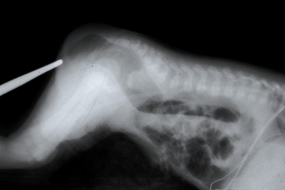
Currarino et al. (1961) and Chamberlain et al. (1965) reported a black family in which 3 sisters had a form of osteoarthropathy seemingly distinct from pachydermoperiostosis. Clinical features included clubbing of the fingers, eczematous skin eruption, increased sweating of the palms and soles, swollen extremities, periosteal new bone formation, and defects of the cranial bones resulting in wide fontanels. Thickened skin was not reported.
Reginato et al. (1982) reported 3 sibs with primary hypertrophic osteoarthropathy and widely open cranial sutures and fontanelles. The syndrome was apparent after birth and included digital clubbing, subperiosteal bone formation, and painful soft tissue swelling over bones. The 2 oldest sibs had almost complete resolution of the cranial defects and bone formation by ages 4 and 6 years, respectively. Joint swelling and clubbing persisted, and mild bone resorption of the distal phalanges became apparent at an older age. Neither parent was affected.
Sinha et al. (1997) reported a boy and girl, both belonging to a large consanguineous Pakistani kindred, with the disorder. The boy presented at age 3 with digital clubbing and extreme hyperhidrosis. Radiographs were not performed. The girl presented at age 11 years with swollen painful knees and hip pain. She had a history of ligation of a patent ductus arteriosus (PDA) and cleft palate repair. There was no skin rash, clubbing of the digits, or excessive sweating. Radiographs of the hands showed soft tissue expansion around the tips of the fingers with irregularities of the distal ends of the terminal phalanges. She developed excessive sweating of the hands a few years later. Eight family members of the patients had digital clubbing; radiographs were not performed. Although Sinha et al. (1997) stated that this family had pachydermoperiostosis, pachydermia was not described, and Dabir et al. (2007) concluded that the family reported by Sinha et al. (1997) had cranioosteoarthropathy, despite the fact that cranial suture abnormalities were not mentioned in the report of Sinha et al. (1997).
O'Connell et al. (2004) reported 2 unrelated Asian children with cranioosteoarthropathy associated with congenital heart disease. The first patient had tetralogy of Fallot complicated by heart block. At age 10 weeks, he was noted to have wide fontanelles with wormian bone formation. Digital clubbing unrelated to heart disease was apparent at 13 months of age. Radiographs at age 19 months showed delayed bone age, periosteal new bone formation along the diaphyses of the long bones, and mild acroosteolysis of the distal phalanges of the fingers. His older sister had a PDA without other features. The second patient, born of consanguineous Asian parents, presented at age 4 years with painful swelling of the knees and ankles, digital clubbing, and excessive sweating of the palms. She had a history of PDA and patent foramen ovale. O'Connell et al. (2004) discussed the difficulties in classification of cranioosteoarthropathy and pachydermoperiostosis, noted the clinical overlap between the 2 conditions, and suggested that they may be allelic disorders. They also suggested that congenital heart defects may be a core features of the expanded phenotype.
Dabir et al. (2007) emphasized that cranioosteoarthropathy is a variant of hypertrophic osteoarthropathy with the additional feature of decreased ossification of the cranium and the absence of pachydermia.
InheritanceRimoin (1965) reported a family with 2 double-consanguineous affected second cousins, indicating autosomal recessive inheritance. Affected persons in successive generations were observed. Recessive inheritance was also suggested by the considerable number of instances of affected sibs with apparently normal parents and the several examples of consanguineous parents (Leva, 1915; Simons, 1918; Shen and Yamanouchi, 1934).
MappingBy genomewide mapping of 2 families with PHO, Uppal et al. (2008) found significant linkage to a 9-Mb region on chromosome 4q33-q34 between markers D4S2979 and D4S415.
Molecular GeneticsUppal et al. (2008) identified 2 different homozygous truncating mutations in the HPGD gene (601688.0002, 601688.0003) in affected members of 2 unrelated families with autosomal recessive primary hypertrophic osteoarthropathy. One of the families had been reported by Latos-Bielenska et al. (2007). Affected individuals from 2 additional families with cranioosteoarthropathy (Sinha et al., 1997; Dabir et al., 2007), had a missense mutation in the HPGD gene (601688.0001). All homozygous affected individuals had significantly increased urinary prostaglandin E2 (PGE2) levels, up to more than 7 times control values. Heterozygous family members had mild digital clubbing that was most apparent in older individuals, suggesting that the carrier state for HPGD mutations results in a modest, chronic elevation of circulating prostaglandin and late-onset clubbing. Four of the 13 HPGD-deficient patients had a persistent patent ductus arteriosus (PDA), which likely resulted from increased PGE2. However, most did not have patent ductus arteriosus, suggesting that HPGD is not absolutely required for ductus closure in humans.
In affected members of 3 unrelated consanguineous Turkish families with primary hypertrophic osteoarthropathy, Yuksel-Konuk et al. (2009) identified homozygosity for missense mutations in the HPGD gene. The A140P mutation (601688.0001), previously detected in 2 Pakistani families with cranioosteoarthropathy by Uppal et al. (2008), was identified in 2 of the families, whereas 3 affected sisters in the third family were homozygous for a missense mutation involving the initiation codon (M1L; 601688.0005).
In a sister and brother from a consanguineous Turkish family with primary hypertrophic osteoarthropathy, Erken et al. (2015) identified homozygosity for a 2-bp deletion in the HPGD gene (601688.0006) that segregated with disease in the family and was not found in 136 Turkish controls. Examination of 7 heterozygous family members revealed no signs of the disease.
Genotype/Phenotype CorrelationsSeifert et al. (2012) observed that in patients with homozygous mutations in the SLCO2A1 gene, manifestations of PHO emerged later than in patients with HPGD mutations, beginning with clubbing of distal phalanges during puberty and pachydermia shortly after puberty. However, the degree of arthritis, joint involvement, and pachydermia in patients with homozygous SLCO2A1 mutations seemed to be more pronounced than in individuals with homozygous or compound heterozygous HPDG mutations.
Diggle et al. (2012) stated that certain clinical features appear characteristic of patients with mutations in HPGD or in SLCO2A1: HPGD-deficient individuals generally present with digital clubbing and other symptoms in early childhood, whereas SLCO2A1-deficient patients are diagnosed later, after puberty or in early adulthood; pachydermia occurs in both groups, but severe cutis gyrata is observed only in the SLCO1A1 group; although periosteal bone deposition is seen in both groups, acroosteolysis is much more prominent in the HPGD-deficient group; and myelofibrosis is associated with biallelic mutations in SLCO2A1 but not HPGD. In regard to the observed excess of males with PHO, Diggle et al. (2012) noted that this had been attributed to higher prostaglandin levels in males, resulting in milder manifestations in females with homozygous mutations in HPGD, and suggested that PHO due to SLCO2A1 mutations might also be a difficult diagnosis to make in females.
HistoryThe first report of digital clubbing is attributed to Hippocrates in the fifth century B.C., and the finding is sometimes referred to as the 'Hippocratic finger' (Coggins et al., 2008; Uppal et al., 2008).