Pneumatosis

Pneumatosis, also known as emphysema, is the abnormal presence of air or other gas within tissues.
In the lungs, emphysema involves enlargement of the distal airspaces, and is a major feature of chronic obstructive pulmonary disease (COPD).
Pneumoperitoneum (or peritoneal emphysema) is air or gas in the abdominal cavity, and is most commonly caused by a perforated abdominal organ. Pneumatosis is also a frequent result of surgery.
Pneumarthrosis, the presence of air in a joint, is rarely a serious sign.
By location
Lungs
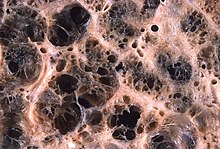
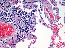
Pulmonary emphysema, more usually called emphysema, is characterised by air-filled cavities or spaces, (pneumatoses) in the lung, that can vary in size and may be very large. The spaces are caused by the breakdown of the walls of the alveoli and they replace the spongy lung parenchyma. This reduces the total alveolar surface available for gas exchange leading to a reduction in oxygen supply for the blood. Emphysema usually affects the middle aged or older population. This is because the disease takes time to develop with the effects of smoking, and other risk factors. Alpha-1 antitrypsin deficiency is a genetic risk factor that may lead to the condition presenting earlier.
It is a typical feature of chronic obstructive pulmonary disease (COPD), a type of obstructive lung disease characterized by long-term breathing problems and poor airflow. Even without COPD, the finding of pulmonary emphysema on a CT lung scan confers a higher mortality in tobacco smokers. In 2016 in the United States there were 6,977 deaths from emphysema – 2.2 per 100,000 of the population. A study on the effects of tobacco and cannabis smoking showed that a possibly cumulative toxic effect could be a risk factor for developing emphysema, and spontaneous pneumothorax. There is an association between emphysema and osteoporosis.
There are three subtypes of pulmonary emphysema – centrilobular or centriacinar, panlobular or panacinar, and paraseptal or distal acinar emphysema, related to the anatomy of the lobules of the lung. These are not associated with fibrosis (scarring). A fourth type known as irregular emphysema involves the acinus irregularly and is associated with fibrosis. Though the subtypes can be seen on imaging they are not well-defined clinically.
Only the first two types of emphysema – centrilobular, and panlobular are associated with significant airflow obstruction, with that of centrilobular emphysema around 20 times more common than panlobular.
Centrilobular emphysema
Centrilobular emphysema also called centriacinar emphysema, affects the centrilobular portion of the lung, the area around the terminal bronchiole, and the first respiratory bronchiole, and can be seen on imaging as an area around the tip of the visible pulmonary artery. Centrilobular emphysema is the most common type usually associated with smoking, and with chronic bronchitis. The disease progresses from the centrilobular portion, leaving the lung parenchyma in the surrounding (perilobular) region preserved. Usually the upper lobes of the lungs are affected.
Panlobular emphysema
Panlobular emphysema also called panacinar emphysema can involve the whole lung or mainly the lower lobes. This type of emphysema is associated with alpha-1 antitrypsin deficiency (A1AD or AATD), and is not related to smoking.
Paraseptal emphysema
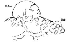
Paraseptal emphysema also called distal acinar emphysema relates to emphysematous change next to a pleural surface, or to a fissure. The cystic spaces known as blebs or bullae that form in paraseptal emphysema typically occur in just one layer beneath the pleura. This distinguishes it from the honeycombing of small cystic spaces seen in fibrosis that typically occurs in layers. This type of emphysema is not associated with airflow obstruction.
Bullous emphysema
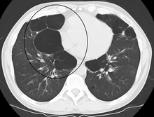
When the subpleural bullae are significant the emphysema is called bullous emphysema. Bullae can become extensive and combine to form giant bullae. These can be large enough to take up a third of a hemithorax, compress the lung parenchyma, and cause displacement. The emphysema is now termed giant bullous emphysema, more commonly called vanishing lung syndrome due to the compressed parenchyma. A bleb or bulla may sometimes rupture and cause a pneumothorax.
HIV associated emphysema
Classic lung diseases are a complication of HIV/AIDS with emphysema being a source of disease. HIV is cited as a risk factor for the development of emphysema regardless of smoking status. HIV associated emphysema occurs over a much shorter time than that associated with smoking. It is thought that an understanding of the mechanisms that underlie the triggering of HIV associated emphysema may help in the understanding of the mechanisms of the development of smoking-related emphysema.
Ritalin lung
The intravenous use of methylphenidate, commonly marketed as Ritalin can lead to emphysematous changes known as Ritalin lung. The mechanism underlying this link is not clearly understood. Ritalin tablets contain talc as a filler, and these need to be crushed and dissolved for injecting. It has been suggested that the talc exposure causes granulomatosis leading to alveolar destruction. However, other intravenous drugs also contain talc and there is no associated emphysematous change. HRCT shows panlobular emphysema.
CPFE
Combined pulmonary fibrosis and emphysema (CPFE) is a rare syndrome that shows upper-lobe emphysema, together with lower-lobe interstitial fibrosis. This is diagnosed by CT scan. This syndrome presents a marked susceptibility for the development of pulmonary hypertension.
Pulmonary interstitial emphysema
Pulmonary interstitial emphysema (PIE) is a collection of air outside of the normal air space of the pulmonary alveoli, found instead inside the connective tissue of the peribronchovascular sheaths, interlobular septa, and visceral pleura.
Congenital lobar emphysema
Congenital lobar emphysema (CLE), also known as congenital lobar overinflation and infantile lobar emphysema, is a neonatal condition associated with enlarged air spaces in the lungs of newborn infants. It is diagnosed around the time of birth or in the first 6 months of life, occurring more often in boys than girls. CLE affects the upper lung lobes more than the lower lobes, and the left lung more often than the right lung. CLE is defined as the hyperinflation of one or more lobes of the lung due to the partial obstruction of the bronchus. This causes symptoms of pressure on the nearby organs. It is associated with several cardiac abnormalities such as patent ductus arteriosus, atrial septal defect, ventricular septal defect, and tetralogy of Fallot. Although CLE may be caused by the abnormal development of bronchi, or compression of airways by nearby tissues, no cause is identified in half of cases. CT scan of the lungs is useful in assessing the anatomy of the lung lobes and status of the neighbouring lobes on whether they are hypoplastic or not. Contrast-enhanced CT is useful in assessing vascular abnormalities and mediastinal masses.
Compensatory emphysema
Compensatory emphysema, is overinflation of part of a lung in response to either removal by surgery of another part of the lung or deceased size of another part of the lung.
Parenchymal focalities

A focal lung pneumatosis, is a solitary volume of air in the lung that is larger than alveoli. A focal lung pneumatosis can be classified by its wall thickness:
- A pulmonary bleb or bulla has a wall thickness of less than 1 mm Blebs and bullae are also known as a focal regions of emphysema.
- A pulmonary cyst has a wall thickness of up to 4 mm. A minimum wall thickness of 1 mm has been suggested, but thin-walled pockets may be included in the definition as well. Pulmonary cysts are not associated with either smoking or emphysema.
- A cavity has a wall thickness of more than 4 mm
The terms above, when referring to sites other than the lungs, often imply fluid content.
Other thoracic
- Pneumothorax, air or gas in the pleural space
- Pneumomediastinum, air or gas in the mediastinum
- Also called mediastinal emphysema or pneumatosis/emphysema of the mediastinum
Abdominal
- Pneumoperitoneum (or peritoneal emphysema), air or gas in the abdominal cavity. The most common cause is a perforated abdominal viscus, generally a perforated peptic ulcer, although any part of the bowel may perforate from a benign ulcer, tumor or abdominal trauma.
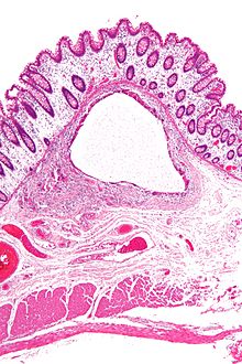
- Pneumatosis intestinalis, air or gas cysts in the bowel wall
- Gastric pneumatosis (or gastric emphysema) is air or gas cysts in the stomach wall
Joints
Pneumarthrosis is the presence of air in a joint. Its presentation on radiography is a radiolucent cleft often called a vacuum phenomenon, or vacuum sign. Pneumarthrosis is associated with osteoarthritis and spondylosis.
Pneumarthrosis is a common normal finding in shoulders as well as in sternoclavicular joints. It is believed to be a cause of the sounds of joint cracking. It is also a common normal post-operative finding at least after spinal surgery. Pneumarthrosis is extremely rare in conjunction with fluid or pus in a joint, and its presence can therefore practically exclude infection.

X-ray of a hip with hip replacement and pneumarthrosis, in this case aseptic.

A vacuum sign, or vacuum phenomenon, is a normal finding on shoulder X-rays.


Other

Subcutaneous emphysema is found in the deepest layer of the skin. Emphysematous cystitis is a condition of gas in the bladder wall. On occasion this may give rise to secondary subcutaneous emphysema which has a poor prognosis.
Pneumoparotitis is the presence of air in the parotid gland caused by raised air pressure in the mouth often as a result of playing wind instruments. In rare cases air may escape from the gland and give rise to subcutaneous emphysema in the face, neck, or mediastinum.
Terminology
The term pneumatosis has word roots of pneumat- + -osis, meaning "air problem/injury".