Breast Cancer

Breast cancer is cancer that develops from breast tissue. Signs of breast cancer may include a lump in the breast, a change in breast shape, dimpling of the skin, fluid coming from the nipple, a newly-inverted nipple, or a red or scaly patch of skin. In those with distant spread of the disease, there may be bone pain, swollen lymph nodes, shortness of breath, or yellow skin.
Risk factors for developing breast cancer include being female, obesity, a lack of physical exercise, alcoholism, hormone replacement therapy during menopause, ionizing radiation, an early age at first menstruation, having children late in life or not at all, older age, having a prior history of breast cancer, and a family history of breast cancer. About 5–10% of cases are the result of a genetic predisposition inherited from a person's parents, including BRCA1 and BRCA2 among others. Breast cancer most commonly develops in cells from the lining of milk ducts and the lobules that supply these ducts with milk. Cancers developing from the ducts are known as ductal carcinomas, while those developing from lobules are known as lobular carcinomas. There are more than 18 other sub-types of breast cancer. Some, such as ductal carcinoma in situ, develop from pre-invasive lesions. The diagnosis of breast cancer is confirmed by taking a biopsy of the concerning tissue. Once the diagnosis is made, further tests are done to determine if the cancer has spread beyond the breast and which treatments are most likely to be effective.
The balance of benefits versus harms of breast cancer screening is controversial. A 2013 Cochrane review found that it was unclear if mammographic screening does more harm than good, in that a large proportion of women who test positive turn out not to have the disease. A 2009 review for the US Preventive Services Task Force found evidence of benefit in those 40 to 70 years of age, and the organization recommends screening every two years in women 50 to 74 years of age. The medications tamoxifen or raloxifene may be used in an effort to prevent breast cancer in those who are at high risk of developing it. Surgical removal of both breasts is another preventive measure in some high risk women. In those who have been diagnosed with cancer, a number of treatments may be used, including surgery, radiation therapy, chemotherapy, hormonal therapy, and targeted therapy. Types of surgery vary from breast-conserving surgery to mastectomy. Breast reconstruction may take place at the time of surgery or at a later date. In those in whom the cancer has spread to other parts of the body, treatments are mostly aimed at improving quality of life and comfort.
Outcomes for breast cancer vary depending on the cancer type, the extent of disease, and the person's age. The five-year survival rates in England and the United States are between 80 and 90%. In developing countries, five-year survival rates are lower. Worldwide, breast cancer is the leading type of cancer in women, accounting for 25% of all cases. In 2018 it resulted in 2 million new cases and 627,000 deaths. It is more common in developed countries and is more than 100 times more common in women than in men.
Signs and symptoms
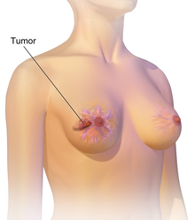

Breast cancer most commonly presents as a lump that feels different from the rest of the breast tissue. More than 80% of cases are discovered when a person detects such a lump with the fingertips. The earliest breast cancers, however, are detected by a mammogram. Lumps found in lymph nodes located in the armpits may also indicate breast cancer.
Indications of breast cancer other than a lump may include thickening different from the other breast tissue, one breast becoming larger or lower, a nipple changing position or shape or becoming inverted, skin puckering or dimpling, a rash on or around a nipple, discharge from nipple/s, constant pain in part of the breast or armpit and swelling beneath the armpit or around the collarbone. Pain ("mastodynia") is an unreliable tool in determining the presence or absence of breast cancer, but may be indicative of other breast health issues.
Another symptom complex of breast cancer is Paget's disease of the breast. This syndrome presents as skin changes resembling eczema; such as redness, discoloration or mild flaking of the nipple skin. As Paget's disease of the breast advances, symptoms may include tingling, itching, increased sensitivity, burning, and pain. There may also be discharge from the nipple. Approximately half the women diagnosed with Paget's disease of the breast also have a lump in the breast.
Inflammatory Breast Cancer presents with similar effects. Inflammatory Breast Cancer is a rare (only seen in less than 5% of breast cancer diagnosis) yet aggressive form of breast cancer characterized by the swollen, red areas formed on the top of the Breast. The visual effects of Inflammatory Breast Cancer is a result of a blockage of lymph vessels by cancer cells. This type of breast cancer is seen in more commonly diagnosed in younger ages, obese women and African American women. As inflammatory breast cancer does not present as a lump there can sometimes be a delay in diagnosis.
In rare cases, what initially appears as a fibroadenoma (hard, movable non-cancerous lump) could in fact be a phyllodes tumor. Phyllodes tumors are formed within the stroma (connective tissue) of the breast and contain glandular as well as stromal tissue. Phyllodes tumors are not staged in the usual sense; they are classified on the basis of their appearance under the microscope as benign, borderline or malignant.
Malignant tumors can result in metastatic tumors— secondary tumors (originating from the primary tumor) that spread beyond the site of origination. The symptoms caused by metastatic breast cancer will depend on the location of metastasis. Common sites of metastasis include bone, liver, lung, and brain. When cancer has reached such an invasive state, it is categorized as a stage 4 cancer, cancers of this state are oftentimes fatal. Common symptoms of stage 4 cancer include unexplained weight loss, bone and joint pain, jaundice and neurological symptoms. These symptoms are called non-specific symptoms because they could be manifestations of many other illnesses. Rarely breast cancer can spread to exceedingly uncommon sites such as peripancreatic lymph nodes causing biliary obstruction leading to diagnostic difficulties.
Most symptoms of breast disorders, including most lumps, do not turn out to represent underlying breast cancer. Less than 20% of lumps, for example, are cancerous, and benign breast diseases such as mastitis and fibroadenoma of the breast are more common causes of breast disorder symptoms.
Risk factors
Risk factors can be divided into two categories:
- modifiable risk factors (things that people can change themselves, such as consumption of alcoholic beverages), and
- fixed risk factors (things that cannot be changed, such as age and biological sex).
The primary risk factors for breast cancer are being female and older age. Other potential risk factors include genetics, lack of childbearing or lack of breastfeeding, higher levels of certain hormones, certain dietary patterns, and obesity. One study indicates that exposure to light pollution is a risk factor for the development of breast cancer.
Lifestyle
Obesity and drinking alcoholic beverages are among the most common modifiable risk factors. However, the correlation between these factors and breast cancer is anything but linear. Studies show that those who rapidly gain weight in adulthood are at higher risk than those who have been overweight since childhood. Likewise excess fat in the midsection seems to induce a higher risk than excess weight carried in the lower body. This implies that the food one eats is of greater importance than one's BMI.
The consumption of alcohol is linked to the risk for breast cancer. Drinking alcoholic beverages increases the risk of breast cancer, even at relatively low (one to three drinks per week) and moderate levels. The risk is highest among heavy drinkers. Dietary factors that may increase risk include a high-fat diet and obesity-related high cholesterol levels. Dietary iodine deficiency may also play a role. Evidence for fiber is unclear. A 2015 review found that studies trying to link fiber intake with breast cancer produced mixed results. In 2016 a tentative association between low fiber intake during adolescence and breast cancer was observed.
Smoking tobacco appears to increase the risk of breast cancer, with the greater the amount smoked and the earlier in life that smoking began, the higher the risk. In those who are long-term smokers, the risk is increased 35% to 50%. A lack of physical activity has been linked to about 10% of cases. Sitting regularly for prolonged periods is associated with higher mortality from breast cancer. The risk is not negated by regular exercise, though it is lowered.
There is an association between use of hormonal birth control and the development of premenopausal breast cancer, but whether birth control pills actually cause premenopausal breast cancer is a matter of debate. If there is indeed a link, the absolute effect is small. Additionally, it is not clear if the association exists with newer hormonal birth controls. In those with mutations in the breast cancer susceptibility genes BRCA1 or BRCA2, or who have a family history of breast cancer, use of modern oral contraceptives does not appear to affect the risk of breast cancer.
The association between breast feeding and breast cancer has not been clearly determined; some studies have found support for an association while others have not. In the 1980s, the abortion–breast cancer hypothesis posited that induced abortion increased the risk of developing breast cancer. This hypothesis was the subject of extensive scientific inquiry, which concluded that neither miscarriages nor abortions are associated with a heightened risk for breast cancer.
Other risk factors include radiation and circadian disruptions related to shift-work and routine late-night eating. A number of chemicals have also been linked, including polychlorinated biphenyls, polycyclic aromatic hydrocarbons, and organic solvents Although the radiation from mammography is a low dose, it is estimated that yearly screening from 40 to 80 years of age will cause approximately 225 cases of fatal breast cancer per million women screened.
Genetics
Genetics is believed to be the primary cause of 5–10% of all cases. Women whose mother was diagnosed before 50 have an increased risk of 1.7 and those whose mother was diagnosed at age 50 or after has an increased risk of 1.4. In those with zero, one or two affected relatives, the risk of breast cancer before the age of 80 is 7.8%, 13.3%, and 21.1% with a subsequent mortality from the disease of 2.3%, 4.2%, and 7.6% respectively. In those with a first degree relative with the disease the risk of breast cancer between the age of 40 and 50 is double that of the general population.
In less than 5% of cases, genetics plays a more significant role by causing a hereditary breast–ovarian cancer syndrome. This includes those who carry the BRCA1 and BRCA2 gene mutation. These mutations account for up to 90% of the total genetic influence with a risk of breast cancer of 60–80% in those affected. Other significant mutations include p53 (Li–Fraumeni syndrome), PTEN (Cowden syndrome), and STK11 (Peutz–Jeghers syndrome), CHEK2, ATM, BRIP1, and PALB2. In 2012, researchers said that there are four genetically distinct types of the breast cancer and that in each type, hallmark genetic changes lead to many cancers.
Other genetic predispositions include the density of the breast tissue and hormonal levels. Women with dense breast tissue are more likely to get tumors and are less likely to be diagnosed with breast cancer - because the dense tissue makes tumors less visible on mammograms. Furthermore, women with naturally high estrogen and progesterone levels are also at higher risk for tumor development.
Medical conditions
Breast changes like atypical ductal hyperplasia and lobular carcinoma in situ, found in benign breast conditions such as fibrocystic breast changes, are correlated with an increased breast cancer risk.
Diabetes mellitus might also increase the risk of breast cancer. Autoimmune diseases such as lupus erythematosus seem also to increase the risk for the acquisition of breast cancer. Hormone therapy to treat menopause is also associated with an increase risk of breast cancer.
Pathophysiology

Breast cancer, like other cancers, occurs because of an interaction between an environmental (external) factor and a genetically susceptible host. Normal cells divide as many times as needed and stop. They attach to other cells and stay in place in tissues. Cells become cancerous when they lose their ability to stop dividing, to attach to other cells, to stay where they belong, and to die at the proper time.
Normal cells will self-destruct (programmed cell death) when they are no longer needed. Until then, cells are protected from programmed death by several protein clusters and pathways. One of the protective pathways is the PI3K/AKT pathway; another is the RAS/MEK/ERK pathway. Sometimes the genes along these protective pathways are mutated in a way that turns them permanently "on", rendering the cell incapable of self-destructing when it is no longer needed. This is one of the steps that causes cancer in combination with other mutations. Normally, the PTEN protein turns off the PI3K/AKT pathway when the cell is ready for programmed cell death. In some breast cancers, the gene for the PTEN protein is mutated, so the PI3K/AKT pathway is stuck in the "on" position, and the cancer cell does not self-destruct.
Mutations that can lead to breast cancer have been experimentally linked to estrogen exposure. Additionally, G-protein coupled estrogen receptors have been associated with various cancers of the female reproductive system including breast cancer.
Abnormal growth factor signaling in the interaction between stromal cells and epithelial cells can facilitate malignant cell growth. In breast adipose tissue, overexpression of leptin leads to increased cell proliferation and cancer.
In the United States, 10 to 20 percent of people with breast cancer and people with ovarian cancer have a first- or second-degree relative with one of these diseases. The familial tendency to develop these cancers is called hereditary breast–ovarian cancer syndrome. The best known of these, the BRCA mutations, confer a lifetime risk of breast cancer of between 60 and 85 percent and a lifetime risk of ovarian cancer of between 15 and 40 percent. Some mutations associated with cancer, such as p53, BRCA1 and BRCA2, occur in mechanisms to correct errors in DNA. These mutations are either inherited or acquired after birth. Presumably, they allow further mutations, which allow uncontrolled division, lack of attachment, and metastasis to distant organs. However, there is strong evidence of residual risk variation that goes well beyond hereditary BRCA gene mutations between carrier families. This is caused by unobserved risk factors. This implicates environmental and other causes as triggers for breast cancers. The inherited mutation in BRCA1 or BRCA2 genes can interfere with repair of DNA cross links and DNA double strand breaks (known functions of the encoded protein). These carcinogens cause DNA damage such as DNA cross links and double strand breaks that often require repairs by pathways containing BRCA1 and BRCA2. However, mutations in BRCA genes account for only 2 to 3 percent of all breast cancers. Levin et al. say that cancer may not be inevitable for all carriers of BRCA1 and BRCA2 mutations. About half of hereditary breast–ovarian cancer syndromes involve unknown genes. Furthermore, certain latent viruses, may decrease the expression of the BRCA1 gene and increase the risk of breast tumors.
GATA-3 directly controls the expression of estrogen receptor (ER) and other genes associated with epithelial differentiation, and the loss of GATA-3 leads to loss of differentiation and poor prognosis due to cancer cell invasion and metastasis.
Diagnosis
Most types of breast cancer are easy to diagnose by microscopic analysis of a sample - or biopsy - of the affected area of the breast. Also, there are types of breast cancer that require specialized lab exams.
The two most commonly used screening methods, physical examination of the breasts by a healthcare provider and mammography, can offer an approximate likelihood that a lump is cancer, and may also detect some other lesions, such as a simple cyst. When these examinations are inconclusive, a healthcare provider can remove a sample of the fluid in the lump for microscopic analysis (a procedure known as fine needle aspiration, or fine needle aspiration and cytology, FNAC) to help establish the diagnosis. A needle aspiration can be performed in a healthcare provider's office or clinic. A local anesthetic may be used to numb the breast tissue to prevent pain during the procedure, but may not be necessary if the lump isn't beneath the skin. A finding of clear fluid makes the lump highly unlikely to be cancerous, but bloody fluid may be sent off for inspection under a microscope for cancerous cells. Together, physical examination of the breasts, mammography, and FNAC can be used to diagnose breast cancer with a good degree of accuracy.
Other options for biopsy include a core biopsy or vacuum-assisted breast biopsy, which are procedures in which a section of the breast lump is removed; or an excisional biopsy, in which the entire lump is removed. Very often the results of physical examination by a healthcare provider, mammography, and additional tests that may be performed in special circumstances (such as imaging by ultrasound or MRI) are sufficient to warrant excisional biopsy as the definitive diagnostic and primary treatment method.


MRI showing breast cancer
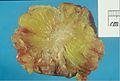
Excised human breast tissue, showing an irregular, dense, white stellate area of cancer 2 cm in diameter, within yellow fatty tissue.
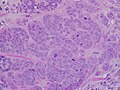
High-grade invasive ductal carcinoma, with minimal tubule formation, marked pleomorphism, and prominent mitoses, 40x field.

Micrograph showing a lymph node invaded by ductal breast carcinoma, with an extension of the tumor beyond the lymph node.
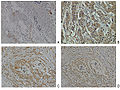
Neuropilin-2 expression in normal breast and breast carcinoma tissue.
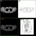
F-18 FDG PET/CT: A breast cancer metastasis to the right scapula

Needle breast biopsy.

Elastography shows stiff cancer tissue on ultrasound imaging.

Ultrasound image shows irregularly shaped mass of breast cancer.

Infiltrating (Invasive) breast carcinoma.


Classification
Breast cancers are classified by several grading systems. Each of these influences the prognosis and can affect treatment response. Description of a breast cancer optimally includes all of these factors.

- Histopathology. Breast cancer is usually classified primarily by its histological appearance. Most breast cancers are derived from the epithelium lining the ducts or lobules, and these cancers are classified as ductal or lobular carcinoma. Carcinoma in situ is growth of low-grade cancerous or precancerous cells within a particular tissue compartment such as the mammary duct without invasion of the surrounding tissue. In contrast, invasive carcinoma does not confine itself to the initial tissue compartment.
- Grade. Grading compares the appearance of the breast cancer cells to the appearance of normal breast tissue. Normal cells in an organ like the breast become differentiated, meaning that they take on specific shapes and forms that reflect their function as part of that organ. Cancerous cells lose that differentiation. In cancer, the cells that would normally line up in an orderly way to make up the milk ducts become disorganized. Cell division becomes uncontrolled. Cell nuclei become less uniform. Pathologists describe cells as well differentiated (low grade), moderately differentiated (intermediate grade), and poorly differentiated (high grade) as the cells progressively lose the features seen in normal breast cells. Poorly differentiated cancers (the ones whose tissue is least like normal breast tissue) have a worse prognosis.
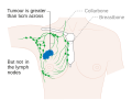
- Stage. Breast cancer staging using the TNM system is based on the size of the tumor (T), whether or not the tumor has spread to the lymph nodes (N) in the armpits, and whether the tumor has metastasized (M) (i.e. spread to a more distant part of the body). Larger size, nodal spread, and metastasis have a larger stage number and a worse prognosis.
The main stages are:- Stage 0 is a pre-cancerous or marker condition, either ductal carcinoma in situ (DCIS) or lobular carcinoma in situ (LCIS).
- Stages 1–3 are within the breast or regional lymph nodes.
- Stage 4 is 'metastatic' cancer that has a less favorable prognosis since it has spread beyond the breast and regional lymph nodes.

Stage T1 breast cancer
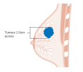
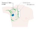
Stage T2 breast cancer
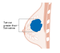
Stage T3 breast cancer

Metastatic or stage 4 breast cancer


- Where available, imaging studies may be employed as part of the staging process in select cases to look for signs of metastatic cancer. However, in cases of breast cancer with low risk for metastasis, the risks associated with PET scans, CT scans, or bone scans outweigh the possible benefits, as these procedures expose the person to a substantial amount of potentially dangerous ionizing radiation.
- Receptor status. Breast cancer cells have receptors on their surface and in their cytoplasm and nucleus. Chemical messengers such as hormones bind to receptors, and this causes changes in the cell. Breast cancer cells may or may not have three important receptors: estrogen receptor (ER), progesterone receptor (PR), and HER2.
ER+ cancer cells (that is, cancer cells that have estrogen receptors) depend on estrogen for their growth, so they can be treated with drugs to block estrogen effects (e.g. tamoxifen), and generally have a better prognosis. Untreated, HER2+ breast cancers are generally more aggressive than HER2- breast cancers, but HER2+ cancer cells respond to drugs such as the monoclonal antibody trastuzumab (in combination with conventional chemotherapy), and this has improved the prognosis significantly. Cells that do not have any of these three receptor types (estrogen receptors, progesterone receptors, or HER2) are called triple-negative, although they frequently do express receptors for other hormones, such as androgen receptor and prolactin receptor. - DNA assays. DNA testing of various types including DNA microarrays have compared normal cells to breast cancer cells. The specific changes in a particular breast cancer can be used to classify the cancer in several ways, and may assist in choosing the most effective treatment for that DNA type.

Stage 1A breast cancer

Stage 1B breast cancer

Stage 2A breast cancer

Stage 2A breast cancer

Stage 2B breast cancer

Stage 2B breast cancer
Stage 2B breast cancer

Stage 3A breast cancer

Stage 3A breast cancer

Stage 3A breast cancer

Stage 3B breast cancer
Stage 3B breast cancer


