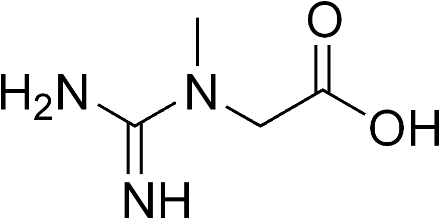
Cerebral Creatine Deficiency Syndrome 3

A number sign (#) is used with this entry because cerebral creatine deficiency syndrome-3 (CCDS3), also known as arginine:glycine amidinotransferase (AGAT) deficiency, is caused by homozygous mutation in the GATM gene (602360) on chromosome 15q21.
DescriptionCerebral creatine deficiency syndrome-3 is an autosomal recessive disorder characterized by developmental delay/regression, mental retardation, severe disturbance of expressive and cognitive speech, and severe depletion of creatine/phosphocreatine in the brain (Schulze, 2003). Most patients develop a myopathy characterized by muscle weakness and atrophy later in life. Oral creatine supplementation can offer symptom improvement (summary by Edvardson et al., 2010).
For a general phenotypic description and a discussion of genetic heterogeneity of cerebral creatine deficiency syndrome, see CCDS1 (300352).
Clinical FeaturesBianchi et al. (2000) reported 2 female sibs, aged 4 and 6 years, with mental retardation and severe creatine deficiency in the brain. GAMT (601240) enzyme activity was normal. Urinary guanidinoacetate concentrations were low, suggesting a deficiency of AGAT. Treatment with oral creatine promptly increased the level of cerebral creatine, which was paralleled by a favorable clinical response, as shown by significant improvement of highly abnormal developmental scores.
Battini et al. (2006) reported an affected 5-year-old cousin of the sisters reported by Bianchi et al. (2000). At age 2 years he presented with psychomotor and language delay and autistic-like behavior. He was found to have severe creatine deficiency in the brain. Creatine monohydrate supplementation normalized creatine levels and resulted in clinical improvement.
Edvardson et al. (2010) reported 2 sibs, born of unrelated Yemenite Jewish parents, with cerebral creatine deficiency syndrome. Both showed delayed psychomotor development and failure to thrive in infancy. At ages 18 and 12 years, respectively, both had mental retardation and decreased muscle mass and strength, mainly proximal, associated with increased serum creatine kinase. Both fatigued easily and refrained from sports. The older sib had high-arched palate, long fingers, and pes cavus. Muscle biopsies showed slight preponderance of type 2 fibers and tubular aggregates. Respiratory chain complex activities were variably decreased. Urinary guanidinoacetate (GAA) levels were significantly decreased, and brain MRS showed decreased creatine signals. Treatment with oral creatine resulted in some clinical improvement and increased cerebral creatine levels.
Verma (2010) reported 2 Jordanian sibs, born of consanguineous parents, with CCDS3. Both showed delayed development in early childhood. At 18 to 20 years, both also developed progressive proximal muscle weakness with features of a myopathy. Muscle biopsy of 1 patient showed fiber size variation and slight atrophy of type 1 and type 2 fibers. Laboratory studies showed undetectable GAA levels. Treatment with oral creatine supplementation resulted in dramatic improvement of muscle strength, but speech and cognitive impairment were unchanged. Genetic analysis identified a homozygous truncating mutation in the GATM gene (R169X; 602360.0003).
Ndika et al. (2012) reported a 9-year-old Chinese girl with CCDS3. She had delayed motor development and hypotonia in infancy and later showed poor overall growth. Laboratory studies yielded generalized organic aciduria, markedly reduced urinary and plasma GAA, and low serum creatine. AGAT enzyme activity was undetectable in cultured lymphoblasts. Early and intense treatment with creatine supplementation resulted in significant developmental progress, with advanced academic performance, but average verbal skills.
Nouioua et al. (2013) reported 2 sisters, aged 11 and 6 years, with CCDS3. They had moderately delayed psychomotor development, language delay, and progressive proximal muscle weakness with Gowers sign and myopathic features on EMG. Laboratory studies showed undetectable guanidinoacetate and low levels of creatine in plasma and urine. Brain MRS showed a markedly reduced level of creatine. Treatment with oral creatine resulted in dramatic improvement in muscle strength and mild improvement in language and cognitive functions. Genetic analysis identified a homozygous missense mutation in the GATM gene (Y203S; 602360.0005).
DiagnosisBattini et al. (2006) noted that low plasma and urine levels of guanidinoacetic acid and creatine levels at birth are indicative of AGAT deficiency. Complete absence of cerebral total creatine by brain proton magnetic spectroscopy confirms the diagnosis.
Among 20 patients referred for genetic testing for mutations in the GATM gene, Comeaux et al. (2013) found that 7 had normal GAA plasma levels and 3 of 6 with initially low GAA levels had normal GAA plasma levels on repeat testing. These 10 patients were not sequenced for GATM mutations. Of the other 10 patients with low GAA levels who were sequenced, only 2 were found to have pathogenic mutations; 8 with low GAA levels had normal GATM sequences. The findings indicated that plasma GAA levels as a biomarker has 100% specificity for GATM mutations (no individuals with normal GAA had mutations), but low sensitivity (low GAA does not necessarily indicate GATM mutations), perhaps due to low GAA levels in normal individuals.
InheritanceThe transmission pattern of AGAT deficiency in the family reported by Bianchi et al. (2000) and Battini et al. (2006) was consistent with autosomal recessive inheritance.
Clinical ManagementBattini et al. (2006) suggested that early treatment in patients with AGAT deficiency may prevent the phenotypic expression of the disease. They treated the affected brother of the sisters reported by Bianchi et al. (2000) with creatine supplementation beginning at age 2 months before the onset of symptoms. At age 18 months, he had normal growth parameters and developmental quotients, whereas his affected relatives, at the same age, already showed a severe delay in somatic growth and psychomotor development, associated with hypotonia and autistic-like behavior.
Molecular GeneticsIn the sibs reported by Bianchi et al. (2000), Item et al. (2001) identified a homozygous mutation in the GATM gene (W149X; 602360.0001). The parents were heterozygous for the mutant allele, with intermediate residual AGAT activities.
In an affected cousin of the sibs reported by Bianchi et al. (2000), Battini et al. (2002) identified homozygosity for the same W149X mutation; his parents and 10 additional subjects in the pedigree were heterozygous for the mutation.
In 2 sibs, born of unrelated Yemenite Jewish parents, with cerebral creatine deficiency syndrome-3, Edvardson et al. (2010) identified a homozygous truncating mutation in the GATM gene (602360.0002).
In a Chinese girl with CCDS3, Ndika et al. (2012) identified a homozygous splice site mutation in the GATM gene (602360.0004).