Juvenile Dermatomyositis

Juvenile dermatomyositis (JDM) is the early-onset form of dermatomyositis (DM, see this term), a systemic, autoimmune inflammatory muscle disorder, characterized by proximal muscle weakness, evocative skin lesion, and systemic manifestations.
Epidemiology
The exact prevalence of JDM is not known. Estimated annual incidence rates range from 1/520,000 to 1/250,000. Females are affected more frequently than males (2.3:1 ratio).
Clinical description
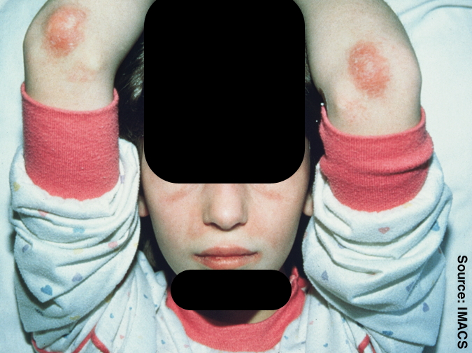
Dermatomyositis occurring before the age of 18 years is considered to be JDM. The average age of onset is 5 to 14 years of age (median = 7 years). Patients commonly have the signs of DM, i.e symmetrical proximal muscle weakness and erythematous rash (heliotrope rash, Gottron papules, sunexposed areas erythema), that is sometimes pruritic, along with cutaneous vasculitis and ulcerations, calcinosis of soft tissue (20-40%), and vasculopathy affecting the digestive tract (with bowel ischemia and/or infarction, abdominal pain, and melena). Muscle weakness leads to variable impairment of physical function. Commonly reported signs also include myalgia and arthralgia. Other associated extramuscular features include dysphagia, sometimes dysphonia, hoarseness, pneumonitis, cardiac manifestations (conduction defects, myocarditis, dilated cardiomyopathy, see this term). Raynaud's phenomenon and inflammatory arthritis. The course of JDM is highly variable: some patients go into remission within 2 to 3 years, others have a cyclic course marked by relapse, while some have ulcerative or chronic disease. Macrophage activation syndrome, a severe sometimes life-threatening condition, has been described in some children diagnosed with JDM. Malignancy and interstitial lung disease are uncommon. Reported malignancies include lymphoma and leukemia.
Etiology
The exact pathogenesis of JDM has not yet been elucidated. It is thought to be related to complement-mediated changes in small vessels in muscle tissue leading to vascular damage. Viruses (e.g. coxsackie virus, parvovirus, and echovirus) and Toxoplasma and Borrelia species, have been suggested as possible triggers.
Diagnostic methods
Diagnosis is based on the clinical signs and magnetic resonance imaging (MRI) of muscle. Muscle biopsy and electromyographic (EMG) testing may also be used but are more invasive. Muscle enzymes are elevated. Nailfold capillaroscopy can be used to predict disease severity and disease course. Antinuclear antibody (ANA), extractable nuclear antigens (ENA); and myositis-specific antibodies (anti-Jo-1, anti-SRP, and anti-Mi-2) should be tested.
Differential diagnosis
Differential diagnosis in JDM may include mitochondrial myopathies, infectious myopathies, other forms of inflammatory myopathies, particularly autoimmune necrotizing myopathy (see this term), as well as Duchenne muscular dystrophy or Becker muscular dystrophy, systemic lupus erythematosus, and juvenile idiopathic arthritis (see these terms).
Management and treatment
The aim of treatment is to reduce long-term morbidity and to restore physical function. High-dose corticosteroids are the mainstay of treatment, with dose tapering after a few weeks of therapy depending on patient response. Methotrexate may also be used, and for severe disease, intravenous methylprednisolone (IVMP). Physical therapy is important to maintain or restore muscle strength. Topical corticosteroids and tacrolimus have been used to treat skin manifestations. Patients should avoid direct UV light and use high-factor sunscreen. No effective treatment is currently available for calcinosis cutis.
Prognosis
Treatment is generally effective, with very low mortality rates reported. The disorder may however be associated with significant morbidity.