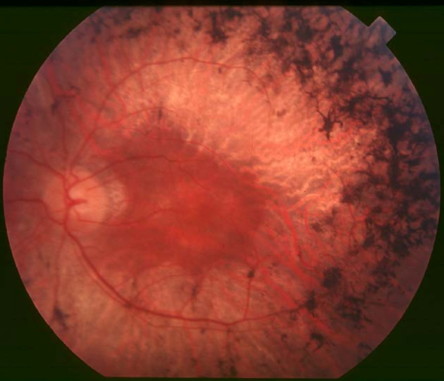
Retinitis Pigmentosa 30

A number sign (#) is used with this entry because of evidence that retinitis pigmentosa-30 (RP30) is caused by mutation in the retinal fascin gene (FSCN2; 607643) on chromosome 17q25.
For a phenotypic description and a discussion of genetic heterogeneity of retinitis pigmentosa, see 268000.
Clinical FeaturesWada et al. (2001) studied 4 Japanese families segregating autosomal dominant retinitis pigmentosa (adRP). All 14 affected patients had had night blindness from childhood. Their visual acuity ranged from hand motion to 1.0. Fundus examination of affected members of 3 generations of 1 family disclosed the progression of retinal degeneration with increasing age. In the early stage, a 10-year-old patient showed a mottled appearance of the retinal pigment epithelium (RPE) and attenuation of the retinal vessels. In all families, affected individuals older than 40 years showed marked retinal degeneration.
Macular Degeneration
Wada et al. (2003) reported 2 Japanese families segregating autosomal dominant macular degeneration. The proband of the first family had gradual progression of visual impairment, photophobia, and constriction of visual fields in his 40s. Examination at 52 years of age showed mottling of the RPE in the posterior pole bilaterally, with diffuse hyperfluorescence around the macula in the right eye and oval and round hyperfluorescent lesions in the posterior pole of the left eye. He also exhibited decreased color vision, but electroretinography (ERG) was within normal limits. He subsequently experienced accelerated deterioration of central visual acuity, reduced to counting fingers bilaterally by age 61. At that time, fundus examination showed bilateral atrophic macular degeneration and a more severely mottled appearance of the RPE, with diffuse retinal mottling in the midperiphery as well as oval areas of hyper- and hypofluorescence within the lesions. Fundus examination of the proband's asymptomatic 37-year-old son showed a mottled appearance of the RPE, yellowish deposits in the macula, and an irregular ring reflex bilaterally. There was granular hyperfluorescence from the posterior pole to the midperiphery bilaterally. In the second family, the 26-year-old proband noted decreased visual acuity in childhood and was diagnosed with retinal degeneration at 8 years of age. Fundus examination at age 13 showed mildly demarcated atrophic macular degeneration associated with diffuse mottling of the retina in the midperiphery bilaterally, with areas of hyper- and hypofluorescence corresponding to RPE atrophy and chorioretinal atrophy, respectively. ERGs showed reduced mixed cone-rod b-wave amplitudes, oscillatory potentials, and 30-Hz flicker responses. He experienced accelerated deterioration of central visual acuity after age 13, and examination at age 24 showed atrophic macular degeneration with progression of pigmentation in both eyes. Kinetic visual field testing revealed absolute scotomas with relatively well-preserved peripheral areas. At 8 years of age, the proband's asymptomatic sister had mottling of the RPE in the posterior poles and reduction of mixed cone-rod a- and b-wave amplitudes as well as oscillatory potentials. At 15 years of age, fundus examination showed tortuous vessels, reddish optic discs, mottling of RPE bilaterally, and pigmentation in the left macula; the atrophic lesions had enlarged slightly since age 8. There was slight peripheral constriction of visual fields but no central or paracentral scotoma. The sibs' asymptomatic 50-year-old mother had tortuosity of retinal vessels and mild RPE atrophy in the posterior poles and midperiphery, as well as mild peripheral constriction of visual fields.
Molecular GeneticsWada et al. (2001) performed mutation screening by SSCP in unrelated Japanese families with RP, including 120 patients with adRP, 200 patients with arRP, and 100 patients with simplex RP. In all 14 affected members from 4 adRP families, Wada et al. (2001) identified a heterozygous 208delG mutation in the retinal FSCN2 gene (607643.0001). The mutation was not found in unaffected members of the 4 families or in any of the autosomal recessive or simplex RP family members. Wada et al. (2001) suggested that the mutation may be relatively common in Japanese patients with adRP.
In 5 affected individuals from 2 Japanese families with macular degeneration, who were negative for mutation in the RDS (PRPH2; 179605), CRX (602225), and GUCY2D (600179) genes, Wada et al. (2003) identified heterozygosity for the same 208delG mutation in the FSCN2 gene (607643.0001) that previously had been identified in patients with autosomal dominant RP (Wada et al., 2001). The mutation segregated with disease in both families.
Gamundi et al. (2005) analyzed the FSCN2 gene in 150 Spanish probands with adRP and 15 with autosomal dominant macular degeneration, 50 patients with sporadic RP, and 50 controls. They detected 16 sequence variations in the patients, including 9 missense mutations, 1 nonsense mutation, and 6 silent mutations, none of which cosegregated with disease in the respective families. Gamundi et al. (2005) suggested that the frequency and type of mutation in FSCN2 might depend on the ethnic population, and proposed that unknown genetic factors might be linked to FSCN2 that could modulate its mutant expression in retinal degeneration.
Zhang et al. (2007) excluded the c.72delG mutation (previously designated 208delG) as a cause of retinal degeneration in a Chinese population. Although the deletion was detected in 8 of 242 patients, including 6 with RP, 1 with Leber congenital amaurosis (see 204000), and 1 with cone-rod dystrophy (see 120970), it was also found in 5 unaffected family members and in 13 of 521 unrelated control subjects. Zhang et al. (2007) cited 3 other studies in which the 72delG mutation was not detected in 458 probands with retinal degeneration from Spanish, Italian, and U.S. populations (Gamundi et al., 2005, Ziviello et al., 2005, Sullivan et al., 2006, respectively). Zhang et al. (2007) also noted that it was highly unusual that the same mutation would cause both rod-cone and cone-rod retinal degeneration.
Jin et al. (2008) studied FSCN2 copy number variation (CNV) and the c.72delG mutation in 54 Japanese controls with a normal retina as well as in 32 patients suspected of having X-linked RP who were negative for mutation in the RPGR (312610) or RP2 (300757) genes. They detected 4 copies of FSCN2 in all individuals, and found the c.72delG mutation in 1 control and in 1 severely affected X-linked RP patient. Analysis of CNV in c.72delG mutation carriers, including 3 previously studied adRP patients and 2 controls, showed that 3 of the RP patients and all 3 controls carrying the c.72delG mutation had a 1:1 ratio of wildtype-to-mutant alleles; however, 1 severely affected RP patient was found to have a 4:1 wildtype-to-mutant allelic ratio. Based on these findings, Jin et al. (2008) suggested that FSCN2 was unlikely to be the pathogenic cause of retinal degeneration.
Animal ModelYokokura et al. (2005) generated mice carrying the Fscn2 208delG mutation or an Fscn2 exon 1-null allele. Homozygotes or heterozygotes for either mutation were morphologically normal, viable, and fertile, but exhibited progressive photoreceptor degeneration with increasing age by light microscopy. Structural abnormalities of the outer segment of the retina were detected by transmission electron microscopy, and electroretinography documented depressed rod and cone responses that also worsened with increasing age.