Retinitis Pigmentosa 69

A number sign (#) is used with this entry because of evidence that retinitis pigmentosa-69 (RP69) is caused by homozygous or compound heterozygous mutation in the KIZ gene (615757) on chromosome 20p11.
DescriptionRetinitis pigmentosa (RP), also designated rod-cone dystrophy, is characterized by initial night blindness due to rod dysfunction, with subsequent progressive constriction of visual fields, abnormal color vision, and eventual loss of central vision due to cone photoreceptor involvement (summary by El Shamieh et al., 2014).
For a discussion of genetic heterogeneity of retinitis pigmentosa, see 268000.
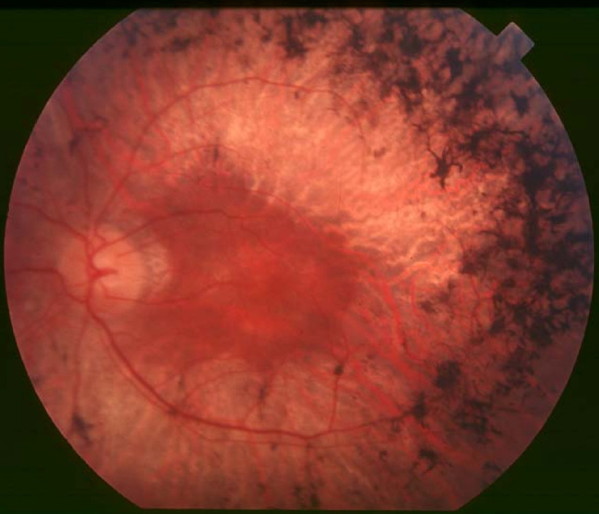
Clinical FeaturesEl Shamieh et al. (2014) studied 3 unrelated patients with rod-cone dystrophy (retinitis pigmentosa) and mutations in the KIZ gene (see MOLECULAR GENETICS). All were diagnosed in their late teens on the basis of night blindness followed by changes in midperipheral visual fields and undetectable responses on full-field electroretinography by approximately 35 years of age. The first was a 50-year-old man of North African Jewish Sephardic descent with unaffected first-cousin parents. Best corrected visual acuity (BCVA) was 20/800 and 20/640 in the right and left eyes, respectively. A kinetic visual field test revealed decreased central retinal sensitivity in addition to bilateral peripheral field constriction. Fundus examination showed typical pigmentary changes of rod-cone dystrophy in the peripheral retina as well as atrophic changes in the central macula with a ring of hypoautofluorescence; spectral-domain optical coherence tomography (OCT) revealed foveal thinning with loss of outer-segment structures. The second patient was a 34-year-old Spanish man whose BCVA was 20/20 in both eyes. Binocular kinetic visual field testing showed an annular scotoma in the midperiphery and preservation of the peripheral isopter, and fundus examination showed mild pigmentary changes in the peripheral retina in association with slight changes in autofluorescence outside the vascular arcades and a perifoveal ring of increased autofluorescence. Foveal structure was normal on OCT, consistent with the patient's normal central vision. The third patient was a 51-year-old man of mixed Italian and French descent whose medical history included congenital ichthyosis (see 242300). BCVA was 20/40 and 20/32 in the right and left eyes, respectively. Visual fields were reduced to the central 10 degrees, with bitemporal islands of perception peripherally. Fundus changes were typical of retinitis pigmentosa, with relative macular preservation. There was a perifoveal ring of increased autofluorescence and moderate thinning of the fovea on OCT.
Molecular GeneticsIn a 50-year-old man of North African Jewish Sephardic descent with retinitis pigmentosa, who was negative for mutation in genes known to be associated with retinal disease, El Shamieh et al. (2014) performed whole-exome sequencing and identified a homozygous nonsense mutation in the KIZ gene (R76X; 615757.0001) that segregated with disease in the family. Analysis of KIZ in 340 unrelated patients with autosomal recessive and sporadic retinitis pigmentosa identified 1 patient who was homozygous for the R76X mutation and another who was compound heterozygous for a different nonsense mutation (E18X; 615757.0002) and a 4-bp deletion (615757.0003). The authors stated that KIZ accounted for about 1% of disease in their cohort of patients, but noted that this might represent a slight overestimation for autosomal recessive retinitis pigmentosa, since in most of their patients mutations in other retinal disease-associated genes had already been excluded.