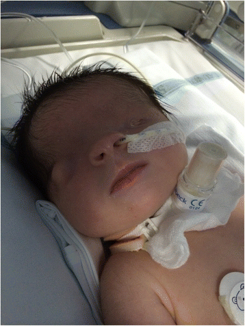
Fraser Syndrome

A rare congenital malformation mainly characterized by unilateral or bilateral cryptophthalmos, syndactyly and urogenital anomalies.
Epidemiology
The estimated prevalence of Fraser syndrome (FS) in Europe is 1/500,000 births. The disorder affects both sexes equally.
Clinical description
FS is usually characterized by eye (unilateral or bilateral cryptophthalmos, microphthalmos/anophthalmos), urinary tract (renal agenesis, bladder atresia/hypoplasia) and genital anomalies (ambiguous genitalia, cryptorchidism, underdevelopment of male genitalia). Cutaneous syndactyly occurs in both hands and feet. Patients may also present craniofacial (dysplastic ears, bifid nose, cleft lip/palate, microglossia), respiratory (laryngeal stenosis/hypoplasia), cardiac (atrial and ventricular septal defect), digestive tract (anal agenesis/imperforation, diaphragmatic hernia, large bowel obstruction) and skeletal anomalies (skull/spine malformations, clubfoot). In case of severe manifestations, the disease may be fatal for the fetus and the major causes of early death after birth include laryngeal and/or kidney anomalies.
Etiology
FS is a genetically heterogeneous disorder, caused by mutations in FRAS1> (4q21.21), FREM2 (13q13.3) and GRIP1 (12q14.3) genes, coding for extracellular matrix proteins essential for the adhesion between basement membrane of epidermis and connective tissues of dermic layer during embryological development. Mutations in these genes are suggested to be responsible for a failure in apoptosis.
Diagnostic methods
The diagnosis of FS is based on clinical findings, imaging and genetic tests for causative mutations. Karyotype, and imaging of urogenital tract may be required in case of ambiguous genitalia. The diagnostic criteria for FS are divided into six major criteria (syndactyly, cryptophthalmos spectrum, urinary tract abnormalities, ambiguous genitalia, laryngeal and tracheal anomalies and a positive family history) and five minor criteria (anorectal defects, dysplastic ears, skull ossification defects, umbilical abnormalities, nasal anomalies). Diagnosis is confirmed in presence of either three major, or two major and two minor, or one major and three minor diagnostic criteria.
Differential diagnosis
The differential diagnoses include ablepharon-macrostomia, fronto-facio-nasal dysplasia, Fraser-like syndrome, Meckel syndrome, and syndromic microphthalmia caused by heterozygous mutations of SOX2 gene. Isolated cryptophthalmos, frontonasal dysplasia should also be considered.
Antenatal diagnosis
Pre-implantation or prenatal genetic diagnosis is possible if pathogenic mutations responsible for the disease have been identified in the family. Polyhydramnios or oligohydramnios, cryptophthalmos, echogenic lungs, renal abnormalities or agenesis may be detected by antenatal ultrasound.
Genetic counseling
FS is an autosomal recessive disease. Genetic counseling should be proposed to at risk families (both parents are carriers of a disease-causing mutation) informing them of the 25% risk of having an affected child at each pregnancy.
Management and treatment
The treatment of FS is symptomatic and requires a multidisciplinary team (including surgeon, ear nose throat (ENT) specialist, nephrologist, ophthalmologist and other specialists). Emergency surgery is required in case of respiratory distress. Patients may also need corrective oculoplastic/corneal, ear, digital or genital surgery. In case of feeding difficulties, a nasogastric tube or gastrostomy may be used, along with physiotherapy to get rid of nasal and oral secretions. Symptomatic treatment of chronic kidney disease may be required. Other treatments may include visual/hearing aids, physiotherapy and psychomotor/occupational/speech therapy. Psychological support should be proposed to the patients and their family.
Prognosis
FS survival depends on the severity of associated anomalies, with laryngeal and/or kidney malformations as the major causes of fatality. The disease can be suspected during the prenatal period by ultrasound detection of related malformations; affected fetuses with severe anomalies are delivered stillborn (about 25%). Nevertheless, in less severe case anomalies, extended or normal life expectancy has been reported.