Malignant Atrophic Papulosis

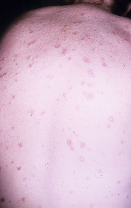
Malignant atrophic papulosis (MAP) is a rare, chronic, thrombo-obliterative vasculopathy characterized by papular skin lesions with central porcelain-white atrophy and a surrounding teleangiectatic rim. Systemic lesions may affect the gastrointestinal tract and the central nervous system (CNS) and are potentially lethal.
Epidemiology
Less than 200 cases have been described in the literature.
Clinical description
MAP onset occurs in adults aged 20-50 with skin lesions that appear initially as small erythematous papules, predominantly on the trunk and the upper extremities. Over several days, the center of the lesions sinks and develops a characteristic morphology: 0.5-1 cm wide papules with an atrophic porcelain-white center and an erythematous, teleangiectatic rim. This condition is chronic and lesions persist over years, often throughout life. Face, scalp, palms of hands and soles of feet are rarely involved. In the systemic variant, which may develop simultaneously or even years after cutaneous symptoms, patients may present with multiple limited infarcts of the intestine, with abdominal pain, bleeding and diarrhea, and/or of the CNS, with cerebrovascular accidents (in rare cases, medullary). It may more rarely manifest as pericarditis or in other organs such as the lungs, presenting as pleuritis. Ocular involvement, which affects the eyelids, conjuctiva, retina, sclera and the choroid plexus, as well as the development of diplopia and ophthalmoplegia (as secondary side effects of the neurologic involvement), has also been described.
Etiology
The etiopathogenesis of the disease remains unknown. Hypotheses implicating vasculitis, coagulopathy or a primary dysfunction of endothelial cells have been proposed. Many patients have been reported to have defects in blood coagulation.
Diagnostic methods
Diagnosis is based primarily on the cutaneous clinical picture that is nearly pathognomonic. In early stages, histology of lesions may reveal a superficial and deep perivascular lymphocytic infiltration with distinct mucin deposition. Later a wedge-shaped connective tissue necrosis in the deep dermis, due to a thrombotic occlusion of the small arteries and sparse lymphocytes, occurs. More developed lesions show prominent changes in the dermoepidermal junction, with atrophy of the epidermis and an area of sclerosis in the papillary dermis.
Differential diagnosis
The histology of early lesions resembles cutaneous lupus erythematosus (see this term). More developed lesions can imitate lichen sclerosus (see this term).
Genetic counseling
A genetic predisposition with an autosomal dominant trait has been suggested.
Management and treatment
Therapeutic efforts with anticoagulants and compounds that facilitate blood perfusion, such as acetylosalicylic acid, pentoxifylline, dipyridamole, ticlodipine and heparin have achieved a partial regression of skin lesions in some individual cases. As all patients may potentially develop the systemic, life-threatening variant, an annual follow-up is mandatory. A clinical inspection of the skin should be combined with additional examinations including brain magnetic resonance tomography, gastroscopy, colonoscopy, X-ray of the chest and abdominal ultrasound, in order to assess the long-term prognosis. No effective treatment for the systemic manifestations has been established, however, subcutaneous treprostinil has been tested successfully in one case with intestinal and CNS manifestations.
Prognosis
Idiopathic, monosymptomatic, cutaneous presentations are benign, however, systemic manifestations can develop years after the occurrence of skin lesions. Systemic manifestations are progressive and may lead to serious complications: bowel perforation and peritonitis as well as thrombosis of the cerebral arteries or massive cerebral hemorrhage, meningitis, encephalitis, radiculopathy and myelitis which have been reported to be lethal in around 50% of patients within 2-3 years.