Retinitis Pigmentosa 83

A number sign (#) is used with this entry because of evidence that retinitis pigmentosa-83 (RP83) is caused by heterozygous mutation in the ARL3 gene (604695) on chromosome 10q24.
For a phenotypic description and a discussion of genetic heterogeneity of retinitis pigmentosa, see 268000.
DescriptionRetinitis pigmentosa-83 (RP83) is characterized by onset of night blindness in the first decade of life, with decreased central vision in the second decade of life in association with retinal degeneration. The retinal dystrophy is associated with cataract, and macular edema has also been reported in some patients (Holtan et al., 2019).
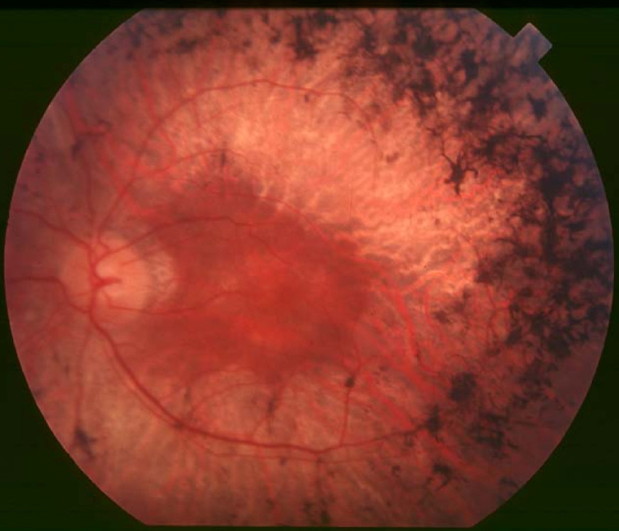
Clinical FeaturesStrom et al. (2016) reported a mother and her son and daughter (pedigree RP02) with retinitis pigmentosa. The 58-year-old mother presented with difficulty reading with decreased visual acuity and bilateral visual field concentric constriction. She had a history of cataract surgery at age 41 years. Visual fields were 'central islands' bilaterally, and the retina showed the classic pattern of mid-peripheral pigmentation with attenuated vessels. Optical coherence tomography (OCT) showed no edema. The 27-year-old daughter had nyctalopia, decreased vision with difficulty reading and driving, flashes and floaters, and progressive loss of peripheral vision. Bilateral visual field constriction was observed. A posterior subcapsular cataract was identified in the left eye, and an intraocular lens in the right eye. Bilateral attenuation of retinal vessels and bone spicules were observed in the periphery. OCT showed cystoid macular edema. The 30-year-old son presented with retinitis pigmentosa and related cystoid macular edema. He also had flashes and floaters, progressive loss of peripheral vision, a posterior subcapsular cataract in the left eye, and an intraocular lens in the right eye.
Holtan et al. (2019) studied a father and son with RP. The 57-year-old Norwegian father had onset of nyctalopia at age 6 years, with narrowing of visual fields in early adulthood, and reduced central vision in the fourth decade of life. The diagnosis of RP was verified by electroretinography (ERG) at age 39, which showed extinguished scotopic and photopic responses. Visual acuity at age 38 was 20/80 bilaterally; by age 52, it was 20/125 to 20/200. Bilateral subcapsular cataracts were removed at ages 46 and 55. Examination at age 57 showed mild asteroid hyalosis in the right vitreous and bilateral severe degeneration of the peripheral and posterior poles of the retina. There was atrophy of the retinal pigment epithelium with bone-spicule pigmentation in the midperiphery, as well as atrophic optic discs and attenuated vessels. He had concentric narrowing of visual fields with 10 degrees of central vision remaining. OCT revealed central atrophy but no cystic macular changes. The proband's son showed pigment changes in the retina at age 16 years and reported some difficulty with night vision but normal peripheral vision. Visual acuities were 20/32 to 20/50. ERG showed extinguished scotopic responses, reduced photopic amplitudes, and increased implicit time, and OCT revealed central atrophy without edema. Examination at age 17 showed a clear lens, no degeneration of the vitreous, and moderate degeneration of the fundus with bone-spicule pigmentation.
InheritanceThe transmission pattern of retinitis pigmentosa in the family reported by Strom et al. (2016) was consistent with autosomal dominant inheritance.
Molecular GeneticsIn a mother and her son and daughter with retinitis pigmentosa, Strom et al. (2016) identified heterozygosity for a missense mutation (Y90C; 604695.0003) in the ARL3 gene. Neither of the mother's parents had the mutation, indicating that it had occurred de novo in the mother. The variant was not present in the Exome Sequencing Project database. No functional studies of the variant were performed.
In a father and son with RP, Holtan et al. (2019) analyzed a panel of 268 retinal disease-associated genes and identified heterozygosity for the Y90C missense mutation in the ARL3 gene, which was shown to have arisen de novo in the father. No ARL3 variants were detected in 431 other patients with eye diseases (primarily RP), suggesting that ARL3 pathogenic variants are a rare cause of RP.