Liver Disease

Liver disease (also called hepatic disease) is a type of damage to or disease of the liver. Whenever the course of the problem lasts long, chronic liver disease ensues.
Signs and symptoms
Some of the signs and symptoms of liver disease are the following:
- Jaundice
- Confusion and altered consciousness caused by hepatic encephalopathy.
- Thrombocytopenia and coagulopathy.
- Risk of bleeding symptoms particularly taking place in gastrointestinal tract
Cause


There are more than a hundred different kinds of liver disease. These are some of the most common:
- Fascioliasis, a parasitic infection of liver caused by a liver fluke of the genus Fasciola, mostly the Fasciola hepatica.
- Hepatitis, inflammation of the liver, is caused by various viruses (viral hepatitis) also by some liver toxins (e.g. alcoholic hepatitis), autoimmunity (autoimmune hepatitis) or hereditary conditions.
- Alcoholic liver disease is a hepatic manifestation of alcohol overconsumption, including fatty liver disease, alcoholic hepatitis, and cirrhosis. Analogous terms such as "drug-induced" or "toxic" liver disease are also used to refer to disorders caused by various drugs.
- Fatty liver disease (hepatic steatosis) is a reversible condition where large vacuoles of triglyceride fat accumulate in liver cells. Non-alcoholic fatty liver disease is a spectrum of disease associated with obesity and metabolic syndrome.
- Hereditary diseases that cause damage to the liver include hemochromatosis, involving accumulation of iron in the body, and Wilson's disease. Liver damage is also a clinical feature of alpha 1-antitrypsin deficiency and glycogen storage disease type II.
- In transthyretin-related hereditary amyloidosis, the liver produces a mutated transthyretin protein which has severe neurodegenerative or cardiopathic effects. Liver transplantation can give a curative treatment option.
- Gilbert's syndrome, a genetic disorder of bilirubin metabolism found in a small percent of the population, can cause mild jaundice.
- Cirrhosis is the formation of fibrous tissue (fibrosis) in the place of liver cells that have died due to a variety of causes, including viral hepatitis, alcohol overconsumption, and other forms of liver toxicity. Cirrhosis causes chronic liver failure.
- Primary liver cancer most commonly manifests as hepatocellular carcinoma or cholangiocarcinoma; rarer forms include angiosarcoma and hemangiosarcoma of the liver. (Many liver malignancies are secondary lesions that have metastasized from primary cancers in the gastrointestinal tract and other organs, such as the kidneys, lungs.)
- Primary biliary cirrhosis is a serious autoimmune disease of the bile capillaries.
- Primary sclerosing cholangitis is a serious chronic inflammatory disease of the bile duct, which is believed to be autoimmune in origin.
- Budd–Chiari syndrome is the clinical picture caused by occlusion of the hepatic vein.
Mechanism
Liver disease can occur through several mechanisms:
DNA damage
One general mechanism, increased DNA damage, is shared by some of the major causes of liver disease. These major causes include infection by hepatitis B virus or hepatitis C virus, alcohol abuse, and obesity.
Viral infection by hepatitis B virus (HBV) or hepatitis C virus (HCV) causes an increase of reactive oxygen species (ROS). The increase in intracellular ROS is about 10,000-fold upon chronic HBV infection and 100,000-fold after HCV infection. This increase in ROS causes inflammation and further increase in ROS. ROS cause more than 20 types of DNA damage. Oxidative DNA damage is mutagenic and also causes epigenetic alterations at the sites of DNA repair. Epigenetic alterations and mutations affect the cellular machinery that may cause the cell to replicate at a higher rate or result in the cell avoiding apoptosis, and thus contribute to liver disease. By the time accumulating epigenetic and mutational changes eventually cause hepatocellular carcinoma, epigenetic alterations appear to have an even larger role in carcinogenesis than mutations. Only one gene, TP53, is mutated in more than 20% of liver cancers while 41 genes each have hypermethylated promoters (repressing gene expression) in more than 20% of liver cancers.
Alcohol consumption in excess causes a build-up of acetaldehyde. Acetaldehyde and free radicals generated by metabolizing alcohol induce DNA damage and oxidative stress. In addition, activation of neutrophils in alcoholic liver disease contributes to the pathogenesis of hepatocellular damage by releasing reactive oxygen species (which can damage DNA). The level of oxidative stress and acetaldehyde-induced DNA adducts due to alcohol consumption does not appear sufficient to cause increased mutagenesis. However, as reviewed by Nishida et al., alcohol exposure, causing oxidative DNA damage (which is repairable), can result in epigenetic alterations at the sites of DNA repair. Alcohol-induced epigenetic alterations of gene expression appear to lead to liver injury and ultimately carcinoma.
Obesity is associated with higher risk of primary liver cancer. As shown with mice, obese mice are prone to liver cancer, likely due to two factors. Obese mice have increased pro-inflammatory cytokines. Obese mice also have higher levels of deoxycholic acid (DCA), a product of bile acid alteration by certain gut microbes, and these microbes are increased with obesity. The excess DCA causes DNA damage and inflammation in the liver, which, in turn, can lead to liver cancer.
Other relevant aspects
A common form of liver disease is viral infection. Viral hepatitides such as Hepatitis B virus and Hepatitis C virus can be vertically transmitted during birth via contact with infected blood. According to a 2012 NICE publication, "about 85% of hepatitis B infections in newborns become chronic". In occult cases, Hepatitis B virus is present by HBV DNA, but testing for HBsAg is negative. High consumption of alcohol can lead to several forms of liver disease including alcoholic hepatitis, alcoholic fatty liver disease, cirrhosis, and liver cancer. In the earlier stages of alcoholic liver disease, fat builds up in the liver's cells due to increased creation of triglycerides and fatty acids and a decreased ability to break down fatty acids. Progression of the disease can lead to liver inflammation from the excess fat in the liver. Scarring in the liver often occurs as the body attempts to heal and extensive scarring can lead to the development of cirrhosis in more advanced stages of the disease. Approximately 3–10% of individuals with cirrhosis develop a form of liver cancer known as hepatocellular carcinoma. According to Tilg, et al., gut microbiome could very well have an effect, be involved in the pathophysiology, on the various types of liver disease which an individual may encounter.
Air pollutants
Particulate matter (PM) or carbon black (CB) are common pollutants. The following factors are the harmful effects of liver exposure under PM or CB. First, they have an obvious direct toxic effect on the liver. Chemicals will affect metabolism and impact liver function. Second, inflammation of liver caused by PM and CB impact lipid metabolism and fatty liver disease. Third, PM and CB can translocate from lung to liver.
PM and CB can easily translocate from lung to liver. Because they are very diverse and each has different toxicodynamics, detailed mechanisms are not clear. Water-soluble fractions of PM is the most important part for PM translocation to liver through extra-pulmonary circulation. When PM goes through blood vessel into blood, it combines with immune cells, that will stimulate innate immune responses. Pro-inflammatory cytokines will be released and cause disease progression.
Diagnosis
A number of liver function tests (LFTs) are available to test the proper function of the liver. These test for the presence of enzymes in blood that are normally most abundant in liver tissue, metabolites or products. serum proteins, serum albumin, serum globulin, alanine transaminase, aspartate transaminase, prothrombin time, partial thromboplastin time.
Imaging tests such as transient elastography, ultrasound and magnetic resonance imaging can be used to examine the liver tissue and the bile ducts. Liver biopsy can be performed to examine liver tissue to distinguish between various conditions; tests such as elastography may reduce the need for biopsy in some situations.
In liver disease, prothrombin time is longer than usual. In addition, the amounts of both coagulation factors and anticoagulation factors are reduced because the liver cannot productively synthsize them as it did when healthy. Nonetheless, there are two exceptions in this falling tendency, that are, coagulation factor VIII and von Willebrand factor, a platelet adhesive protein. Both inversely rise in the setting of hepatic insufficiency, thanks to the drop of hepatic clearance and compensatory productions from other sites of the body. Fibrinolysis generally proceeds faster in the scenarios of acute liver failure as well as advanced stage of liver disease in contrast to chronic liver disease in which concentration of fibrinogen remains unchanged.
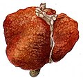
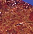
A previously undiagnosed liver disease may become evident first after autopsy. Following are gross pathology images:

Diffuse cirrhosis

Macronodular cirrhosis

Nutmeg texture of congestive hepatopathy

Liver metastases
Treatment

Anti-viral medications are available to treat infections such as hepatitis B. Other conditions may be managed by slowing down disease progression, for example:
- By using steroid-based drugs in autoimmune hepatitis.
- Regularly removing a quantity of blood from a vein (venesection) in the iron overload condition, hemochromatosis.
- Wilson’s disease, a condition where copper builds up in the body, can be managed with drugs that bind copper, allowing it to be passed from the body in urine.
- In cholestatic liver disease, (where the flow of bile is affected due to cystic fibrosis) a medication called ursodeoxycholic acid (URSO, also referred to as UDCA) may be given.
See also
- Model for end-stage liver disease (MELD)


