Retinitis Pigmentosa 20

A number sign (#) is used with this entry because autosomal recessive retinitis pigmentosa-20 (RP20) is caused by homozygous or compound heterozygous mutation in the RPE65 gene (180069) on chromosome 1p31.
Mutations in the RPE65 gene also cause Leber congenital amaurosis (LCA2; 204100).
For a phenotypic description and a discussion of genetic heterogeneity of retinitis pigmentosa (RP), see 268000.
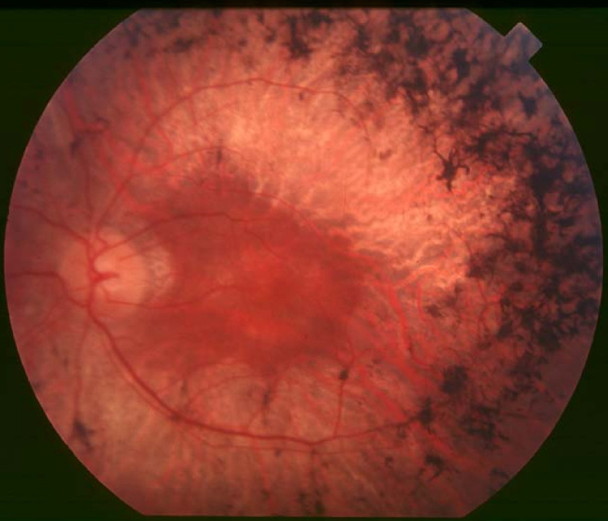
Clinical FeaturesGu et al. (1997) described a 5-generation consanguineous Indian family with 4 members with childhood-onset severe retinal dystrophy (RP20). Onset of severe visual impairment was between 3 and 7 years of age. Night blindness was a typical and early symptom in all patients. Two patients had nystagmus. Ophthalmoscopy revealed attenuated vessels and atrophy of the optic disc. Although bone spicule formation is not a typical feature of the disease, many whitish dots seen on ophthalmoscopy were considered compatible with extensive retinal pigment epithelium (RPE) defects.
In a screen of the RPE65 gene in 147 unrelated patients with autosomal recessive RP and 15 patients with isolated RP, Morimura et al. (1998) identified 3 patients with mutation in RPE65. In one family, from the Dominican Republic, 3 branches of the family had affected children due to homozygosity or compound heterozygosity.
Among 59 probands with RP, 11 with autosomal recessive inheritance, Kondo et al. (2004) identified 1 patient with RP20 (mutation in the RPE65 gene). This 55-year-old Japanese woman, the child of first-cousin parents, had been diagnosed with RP at the age of 40. She had observed the development of night blindness in early childhood and had been free from visual disability until 24 years of age. At the age of 54, she had only basic light-dark perception in both eyes. An examination of the fundus revealed pigmented lesions in the form of clumps or bony spicules involving the posterior retina and associated with a wide area of chorioretinal atrophy, which was prominent in the peripapillary area in both eyes. Electroretinogram showed no recordable rod or cone response in either eye.
Kondo et al. (2004) summarized the effect of RPE65 mutation. The most common phenotype is severe and early-onset retinal degeneration. In most patients with RPE65 mutation, disease was diagnosed in infancy, with visual impairment associated with nystagmus, night blindness, and a tendency to fixate on light. In contrast, the visual performance of several patients in bright light was sufficient to permit attendance at regular school during the elementary years. At older ages, often during the secondary school years, visual acuity was greatly reduced.
Morimura et al. (1998) summarized the clinical criteria distinguishing RP from Leber congenital amaurosis (LCA). RP is the diagnosis given to patients with photoreceptor degeneration who have good central vision within the first decade of life. The diagnosis of LCA is given to patients who are born blind or who lose vision within a few months after birth. Both diagnostic entities feature attenuated retinal vessels and a variable amount of retinal pigmentation in older patients and a reduced or nondetectable electroretinogram at all ages. There is no universally accepted diagnostic term for those patients with retinal degeneration who lose useful (ambulatory) vision during the first few years of life, with ophthalmologists considering such cases as either LCA or severe RP.
MappingGu et al. (1997) used homozygosity mapping in a consanguineous Indian family with 4 affected individuals to map the RP20 locus to chromosome 1p22-p31.
Molecular GeneticsGu et al. (1997) identified 5 families with autosomal recessive childhood-onset severe retinal dystrophy and mutations in the RPE65 gene. Five presumed pathogenic RPE65 mutations (e.g., 180069.0003) were found on a total of 9 alleles in 5 probands. Gu et al. (1997) gave the approximate frequency of RPE65-associated autosomal recessive CSRD as 5%, about the same as for other retinal dystrophy genes. The autosomal recessive mode of inheritance and the 4 potentially inactivating mutations suggested that mutations in RPE65 result in complete or partial loss of protein function.
Among 162 unrelated patients with recessive or sporadic RP, Morimura et al. (1998) identified 3 probands with homozygous or compound heterozygous mutation in the RPE65 gene, 2 with recessive RP (e.g., 180069.0004) and 1 with sporadic RP recategorized as recessive (see 180069.0006). Based on their results, Morimura et al. (1998) estimated that mutations in the RPE65 gene account for approximately 2% of cases of recessive RP.
Kondo et al. (2004) identified a homozygous missense mutation in the RPE65 gene (180069.0008) in a 55-year-old Japanese woman with RP. The authors noted that this mutation had previously been found to result in LCA.