Retinitis Pigmentosa 34

For a phenotypic description and a discussion of genetic heterogeneity of retinitis pigmentosa, see 268000.
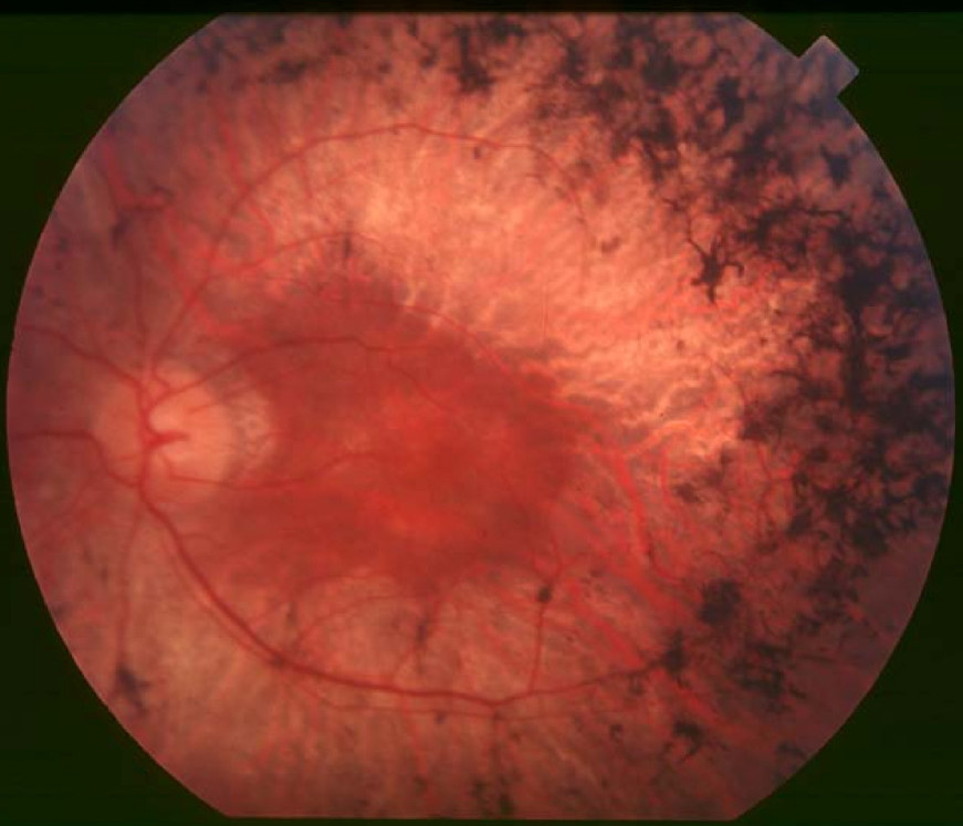
Clinical FeaturesMelamud et al. (2006) described a 3-generation family in which retinitis pigmentosa appeared to be transmitted in an X-linked manner. Four affected individuals who were examined described onset of night blindness, and some described color vision difficulties in the second decade of life, with the earliest at 13 years of age. Indirect ophthalmoscopy revealed peripheral bone spicules, waxy pallor of the optic nerve, and attenuated retinal blood vessels in all patients. All had evidence of constricted peripheral vision. Visual acuity ranged from 20/40 to 20/80+. Based on full-field electroretinography, cone function was more severely reduced than rod function. Female carriers had no ocular signs or symptoms and slightly reduced cone electroretinographic responses.
MappingBy genotyping and linkage analyses of a 3-generation family with retinitis pigmentosa, Melamud et al. (2006) identified significant linkage to markers in the Xq28 region. Mutation analysis of 2 genes within the critical region, CNGA2 (300338) and MPP1 (305360), showed no disease-causing mutations. Linkage analysis indicated that this locus was distinct from that of RP24 (300155), which maps to Xq26-q27. Melamud et al. (2006) raised the possibility that the family had an unusual type of cone-rod dystrophy and the possibility that this represented CORDX2 (300085), the gene locus of which may overlap with the locus defined by their family. However, patients with CORDX2 have a number of clinical features that distinguish them from the affected members of this family. In particular, patients with CORDX2 have early-onset photophobia, color vision defects, and macular atrophic changes.