Deep Vein Thrombosis

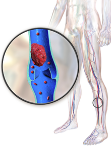
Deep vein thrombosis (DVT) is the formation of a blood clot in a deep vein, most commonly in the legs or pelvis. Symptoms can include pain, swelling, redness, and enlarged veins in the affected area, but some DVTs have no symptoms. The most common life-threatening concern with DVT is the potential for a clot (or multiple clots) to detach from the veins (embolize), travel through the right side of the heart, and become stuck in arteries that supply blood to the lungs. This is called pulmonary embolism (PE). Both DVT and PE are considered as part of the same overall disease process, which is called venous thromboembolism (VTE). VTE can occur as DVT only, as PE with DVT, or PE without DVT. The most frequent long-term complication is post-thrombotic syndrome, which can cause pain, swelling, a sensation of heaviness, itching, and in severe cases, ulcers. Also, recurrent VTE occurs in about 30% of those in the ten years following an initial VTE.
The mechanism of clot formation typically involves some combination of decreased blood flow rate, increased tendency to clot, and injury to the blood vessel wall. Risk factors include recent surgery, older age, active cancer, obesity, personal history and family history of VTE, trauma, injuries, lack of movement, hormonal birth control, pregnancy and the period following birth, and antiphospholipid syndrome. VTE has a strong genetic component, accounting for approximately 50 to 60% of the variability in VTE rates. Genetic factors include non-O blood type, deficiencies of antithrombin, protein C, and protein S and the mutations of factor V Leiden and prothrombin G20210A. In total, dozens of genetic risk factors have been identified.
People suspected of having DVT can be assessed using a prediction rule such as the Wells score. A D-dimer test can also be used to assist with excluding the diagnosis or to signal a need for further testing. Diagnosis is most commonly confirmed by ultrasound of the suspected veins. An estimated 4–10% of DVTs affect the arms. About 5–11% of people will develop VTE in their lifetime, with VTE becoming much more common with age. When compared to those aged 40 and below, people aged 65 and above are at an approximate 15 times higher risk. However, available data has been historically dominated by European and North American populations, and Asian and Hispanic individuals have a lower VTE risk than whites or Blacks.
Using blood thinners (anticoagulation) is the standard treatment, and typical medications include rivaroxaban, apixaban, and warfarin. Beginning warfarin treatment requires an additional non-oral anticoagulant, often injections of heparin. Prevention of VTE for the general population includes avoiding obesity and maintaining an active lifestyle. Preventive efforts following low-risk surgery include early and frequent walking. Riskier surgeries generally prevent VTE with a blood thinner or aspirin combined with intermittent pneumatic compression.
Background
Blood has a natural propensity to clot when blood vessels are damaged (hemostasis) to minimize blood loss. Clotting is activated by the coagulation cascade and the clearing of clots that are no longer needed is accomplished by the fibrinolytic system or fibrinolysis. Reductions in fibrinolysis or increases in coagulation can increase the risk of DVT.
The most common cause of death associated with DVT is when a blood clot (or multiple clots) detach from the veins (embolize), travel through the right side of the heart, and become stuck in pulmonary arteries that supply deoxygenated blood to the lungs for oxygenation. This blockage of blood flow to the lungs is called pulmonary embolism (PE). PE is most common in larger DVTs that occur in the thigh or pelvis. Both DVT and PE are considered as part of the same overall disease process, venous thromboembolism (VTE), which can occur as DVT or PE with or without DVT. VTE is the third most common cause of death from cardiovascular disease, with the top two causes being coronary heart disease and ischemic stroke. In addition to PE, another life-threatening concern with DVT, albeit rare, is when severe cases completely block the venous outflow of a region of the body. This can cause increased pressure leading to compartment syndrome and decreased oxygenation leading to gangrene.
Signs and symptoms
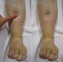
Signs and symptoms of DVT, while highly variable, include pain or tenderness, swelling, warmth, dilation of surface veins, redness or discoloration, and cyanosis with fever. However, some with DVT have no symptoms. Signs and symptoms alone are not sufficiently sensitive or specific to make a diagnosis, but when considered in conjunction with pre-test probability, can help determine the likelihood of DVT. In most suspected cases, DVT is ruled out after evaluation, and symptoms are more often due to other causes, such as ruptured Baker's cyst, cellulitis, hematoma, lymphedema, and chronic venous insufficiency. Other differential diagnoses include tumors, venous or arterial aneurysms, and connective tissue disorders.
Causes
The three factors of Virchow's triad—venous stasis, hypercoagulability, and changes in the endothelial blood vessel lining—contribute to VTE and are used to explain its formation. Venous stasis is the most consequential of these three factors. Other related causes include activation of immune system components, the state of microparticles in the blood, the concentration of oxygen, and possible platelet activation. Various risk factors contribute to VTE, including genetic and environmental factors, though many with multiple risk factors never develop it.
Acquired risk factors include the strong risk factor of older age, which alters blood composition to favor clotting. Previous VTE, particularly unprovoked VTE, is a strong risk factor. Major surgery and trauma increase risk because of tissue factor from outside the vascular system entering the blood. Minor injuries, lower limb amputation, hip fracture, and long bone fractures are also risks. In orthopedic surgery, venous stasis can be temporarily provoked by a cessation of blood flow as part of the procedure. Inactivity and immobilization contribute to venous stasis, as with orthopedic casts, paralysis, sitting, long-haul travel, bed rest, hospitalization, and in survivors of acute stroke. Conditions that involve compromised blood flow in the veins are May–Thurner syndrome, where a vein of the pelvis is compressed, and venous thoracic outlet syndrome, which includes Paget–Schroetter syndrome, where compression occurs near the base of the neck.
Cancer can grow in and around veins, causing venous stasis, and can also stimulate increased levels of tissue factor. Cancers of the bone, ovary, brain, pancreas, and lymphomas are especially associated with increased VTE risk. Chemotherapy treatment also increases risk. Obesity increases the potential of blood to clot, as does pregnancy. In the postpartum, placental tearing releases substances that favor clotting. Oral contraceptives and hormonal replacement therapy increase the risk through a variety of mechanisms, including altered blood coagulation protein levels and reduced fibrinolysis.

Genetic factors account for approximately 50 to 60% of the variability in VTE rates. Family history of VTE is a risk factor for a first VTE. Genetic factors that increase the risk of VTE include deficiencies of three proteins that normally prevent blood from clotting—protein C, protein S, and antithrombin. Deficiencies in antithrombin, protein C, and protein S are rare but strong, or moderately strong, risk factors. These three deficiencies increase the risk of VTE by about 10 times. Factor V Leiden, which makes factor V resistant to inactivation by activated protein C, mildly increases VTE risk by about three times. Having a non-O blood type roughly doubles VTE risk. Non-O blood type is common globally, making it an important risk factor. Individuals without O blood type have higher blood levels of von Willebrand factor and factor VIII than those with O blood type, increasing the likelihood of clotting. Those homozygous for the common fibrinogen gamma gene variant rs2066865 have about a 1.6 times higher risk of VTE. The genetic variant prothrombin G20210A, which increases prothrombin levels, increases risk by about 2.5 times. Additionally, approximately 5% of people have been identified with a background genetic risk comparable to the factor V Leiden and prothrombin G20210A mutations.
Blood alterations including dysfibrinogenemia, low free protein S, activated protein C resistance, homocystinuria, hyperhomocysteinemia, high fibrinogen levels, high factor IX levels, and high factor XI levels are associated with increased risk. Infection, including that of COVID-19, increases risk. Inflammatory diseases such as Behçet's syndrome, and some autoimmune diseases, such as primary antiphospholipid syndrome and systemic lupus erythematosus (SLE), increase risk. SLE itself is frequently associated with secondary antiphospholipid syndrome. Other associated conditions include heparin-induced thrombocytopenia, thrombotic storm, catastrophic antiphospholipid syndrome, paroxysmal nocturnal hemoglobinuria, nephrotic syndrome, chronic kidney disease, HIV, polycythemia vera, intravenous drug use, and smoking.
Some risk factors influence the location of DVT within the body. In isolated distal DVT, the profile of risk factors appears distinct from proximal DVT. Transient factors, such as surgery and immobilization, appear to dominate, whereas thrombophilias and age do not seem to increase risk. Common risk factors for having an upper extremity DVT include having an existing foreign body (such as a central venous catheter, a pacemaker, or a triple-lumen PICC line), cancer, and recent surgery.
Classification

Provoked DVTs occur in association with acquired risk factors, such as surgery, oral contraceptives, trauma, immobility, obesity, or cancer; cases without acquired states are called unprovoked or idiopathic. Acute DVT is characterized by pain and swelling and is usually occlusive, which means that it obstructs blood flow, whereas non-occlusive DVT is less symptomatic. The label "chronic" has been applied to symptomatic DVT that persists longer than 10 to 14 days. DVT that has no symptoms, but is found only by screening, is labeled asymptomatic or incidental. An initial episode of DVT is called incident and any subsequent DVT is termed recurrent. Bilateral DVT refers to clots in both legs while unilateral means that only a single leg is affected.
DVT in the legs is proximal when above the knee and distal (or calf) when below the knee. DVT below the popliteal vein, a proximal vein behind the knee, is classified as distal and has limited clinical significance compared to proximal DVT. Iliofemoral DVT has been described as involving either the iliac or common femoral vein; elsewhere, it has been defined as involving at a minimum the common iliac vein, which is near the top of the pelvis. Upper extremity DVT occurs in the arms or the base of the neck. A rare and massive DVT that causes significant obstruction is phlegmasia cerulea dolens, so named because of observed cases with a blue or purplish discoloration. It is a particularly severe form of acute, proximal, and occlusive DVT. It is life-threatening, limb-threatening, and carries a risk of venous gangrene. It can occur in the arm but more commonly affects the leg.
Pathophysiology

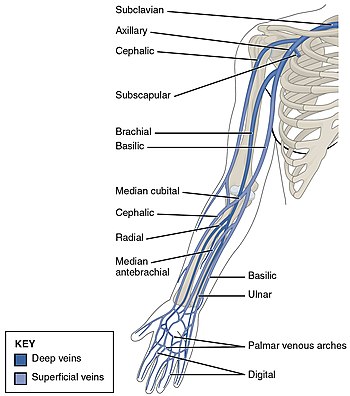
DVT often develops in the calf veins and "grows" in the direction of venous flow, towards the heart. When DVT does not grow, it can be cleared naturally and dissolved into the blood (fibrinolysis). Veins in the leg or pelvis are most commonly affected, including the popliteal vein (behind the knee), femoral vein (of the thigh), and iliac veins of the pelvis. Extensive lower-extremity DVT can even reach into the inferior vena cava (in the abdomen). Upper extremity DVT most commonly affects the subclavian, axillary, and jugular veins.
The causes of arterial thrombosis, such as with heart attacks, are more clearly understood than those of venous thrombosis. With arterial thrombosis, blood vessel wall damage is required, as it initiates coagulation, but clotting in the veins mostly occurs without any such damage. The beginning of venous thrombosis is thought to be caused by tissue factor, which leads to conversion of prothrombin to thrombin, followed by fibrin deposition. Red blood cells and fibrin are the main components of venous thrombi, and the fibrin appears to attach to the blood vessel wall lining (endothelium), a surface that normally acts to prevent clotting. Platelets and white blood cells are also components. Platelets are not as prominent in venous clots as they are in arterial ones, but they can play a role. Inflammation is associated with VTE, and white blood cells play a role in the formation and resolution of venous clots.

Often, DVT begins in the valves of veins. The blood flow pattern in the valves can cause low oxygen concentrations in the blood (hypoxemia) of a valve sinus. Hypoxemia, which is worsened by venous stasis, activates pathways—ones that include hypoxia-inducible factor-1 and early-growth-response protein 1. Hypoxemia also results in the production of reactive oxygen species, which can activate these pathways, as well as nuclear factor-κB, which regulates hypoxia-inducible factor-1 transcription. Hypoxia-inducible factor-1 and early-growth-response protein 1 contribute to monocyte association with endothelial proteins, such as P-selectin, prompting monocytes to release tissue factor-filled microvesicles, which presumably begin clotting after binding to the endothelial surface.
D-dimers are a fibrin degradation product, a natural byproduct of fibrinolysis that is typically found in the blood. An elevated level can result from plasmin dissolving a clot—or other conditions. Hospitalized patients often have elevated levels for multiple reasons. Anticoagulation, the standard treatment for DVT, prevents further clot growth and PE, but does not act directly on existing clots.
Diagnosis
A clinical probability assessment using the Wells score (see dedicated column in the table below) to determine if a potential DVT is "likely" or "unlikely" is typically the first step of the diagnostic process. The score is used in suspected first lower extremity DVT (without any PE symptoms) in primary care and outpatient settings, including the emergency department. The numerical result (possible score −2 to 9) is most commonly grouped into either "unlikely" or "likely" categories. A Wells score of two or more means DVT is considered "likely" (about a 28% chance), while those with a lower score are considered "unlikely" to have DVT (about a 6% chance). In those unlikely to have DVT, a diagnosis is excluded by a negative D-dimer blood test. In people with likely DVT, ultrasound is the standard imaging used to confirm or exclude a diagnosis. Imaging is also needed for hospital inpatients with suspected DVT and those initially categorized as unlikely to have DVT but who have a positive D-dimer test.
While the Wells score is the predominant and most studied clinical prediction rule for DVT, it does have drawbacks. The Wells score requires a subjective assessment regarding the likelihood of an alternate diagnosis and performs less well in the elderly and those with a prior DVT. The Dutch Primary Care Rule has also been validated for use. It contains only objective criteria but requires obtaining a D-dimer value. With this prediction rule, three points or less means a person is at low risk for DVT. A result of four or more points indicates an ultrasound is needed. Instead of using a prediction rule, experienced physicians can make a pre-test DVT probability assessment using clinical assessment and gestalt, but prediction rules are more reliable.
| Criteria | Wells score for DVT | Dutch Primary Care Rule |
|---|---|---|
| Active cancer (treatment within last 6 months or palliative) | +1 point | +1 point |
| Calf swelling ≥ 3 cm compared to asymptomatic calf (measured 10 cm below tibial tuberosity) | +1 point | +2 points |
| Swollen unilateral superficial veins (non-varicose, in symptomatic leg) | +1 point | +1 point |
| Unilateral pitting edema (in symptomatic leg) | +1 point | — |
| Previous documented DVT | +1 point | — |
| Swelling of entire leg | +1 point | — |
| Localized tenderness along the deep venous system | +1 point | — |
| Paralysis, paresis, or recent cast immobilization of lower extremities | +1 point | — |
| Recently bedridden ≥ 3 days, or major surgery requiring regional or general anesthetic in the past 12 weeks | +1 point | +1 point |
| Alternative diagnosis at least as likely | −2 points | — |
| Positive D-dimer (≥ 0.5 mcg/mL or 1.7 nmol/L) | — | +6 points |
| Absence of leg trauma | — | +1 point |
| Male sex | — | +1 point |
| Use of oral contraceptives | — | +1 point |
Compression ultrasonography for suspected deep vein thrombosis is the standard diagnostic method, and it is highly sensitive for detecting an initial DVT. A compression ultrasound is considered positive when the vein walls of normally compressible veins do not collapse under gentle pressure. Clot visualization is sometimes possible, but is not required. Three compression ultrasound scanning techniques can be used, with two of the three methods requiring a second ultrasound some days later to rule out the diagnosis. Whole-leg ultrasound is the option that does not require a repeat ultrasound, but proximal compression ultrasound is frequently used because distal DVT is only rarely clinically significant. Ultrasound methods including duplex and color flow Doppler can be used to further characterize the clot and Doppler ultrasound is especially helpful in the non-compressible iliac veins.
CT scan venography, MRI venography, or a non-contrast MRI are also diagnostic possibilities. The gold standard for judging imaging methods is contrast venography, which involves injecting a peripheral vein of the affected limb with a contrast agent and taking X-rays, to reveal whether the venous supply has been obstructed. Because of its cost, invasiveness, availability, and other limitations, this test is rarely performed.

An ultrasound with a blood clot visible in the left common femoral vein. (The common femoral vein is distal to the external iliac vein.)

Doppler ultrasonography showing absence of flow and hyperechogenic content in a clotted femoral vein (labeled subsartorial) distal to the branching point of the deep femoral vein. When compared to this clot, clots that instead obstruct the common femoral vein cause more severe effects due to impacting a significantly larger portion of the leg.

An abdominal CT scan demonstrating an iliofemoral DVT, with the clot in the right common iliac vein of the pelvis


Management
Treatment for DVT is warranted when the clots are either proximal, distal and symptomatic, or upper extremity and symptomatic. Providing anticoagulation, or blood-thinning medicine, is the typical treatment after patients are checked to make sure they are not subject to bleeding. However, treatment varies depending upon the location of DVT. For example, in cases of isolated distal DVT, ultrasound surveillance (a second ultrasound after 2 weeks to check for proximal clots), might be used instead of anticoagulation. Although, those with isolated distal DVT at a high-risk of VTE recurrence are typically anticoagulated as if they had proximal DVT. Those at a low-risk for recurrence might receive a four to six week course of anticoagulation, lower doses, or no anticoagulation at all. In contrast, those with proximal DVT should receive at least 3 months of anticoagulation.
Some anticoagulants can be taken by mouth, and these oral medicines include warfarin (a vitamin K antagonist), rivaroxaban (a factor Xa inhibitor), apixaban (a factor Xa inhibitor), dabigatran (a direct thrombin inhibitor), and edoxaban (a factor Xa inhibitor). Other anticoagulants cannot be taken by mouth. These parenteral (non-oral) medicines include low-molecular-weight heparin, fondaparinux, and unfractionated heparin. Some oral medicines are sufficient when taken alone, while others require the use of an additional parenteral blood thinner. Rivaroxaban and apixaban are the typical first-line medicines, and they are sufficient when taken orally. Rivaroxaban is taken once daily, and apixaban is taken twice daily. Warfarin, dabigatran, and edoxaban require the use of a parenteral anticoagulant to initiate oral anticoagulant therapy. When warfarin is initiated for VTE treatment, a 5-day minimum of a parenteral anticoagulant together with warfarin is given, which is followed by warfarin only therapy. Warfarin is taken to maintain an international normalized ratio (INR) of 2.0–3.0, with 2.5 as the target. The benefit of taking warfarin declines as the duration of treatment extends, and the risk of bleeding increases with age. Periodic INR monitoring is not necessary when first-line direct oral anticoagulants are used. Overall, anticoagulation therapy is complex and many circumstances can affect how these therapies are managed.


The duration of anticoagulation therapy (whether it will last 4 to 6 weeks, 6 to 12 weeks, 3 to 6 months, or indefinitely) is a key factor in clinical decision making. When proximal DVT is provoked by surgery or trauma a 3-month course of anticoagulation is standard. When a first VTE is proximal DVT that is either unprovoked or associated with transient non-surgical risk factor, low-dose anticoagulation beyond 3 to 6 months might be used. In those with an annual risk of VTE in excess of 9%, as after an unprovoked episode, extended anticoagulation is a possibility. Those who finish warfarin treatment after idiopathic VTE with an elevated D-dimer level show an increased risk of recurrent VTE (about 9% vs about 4% for normal results), and this result might be used in clinical decision making. Thrombophilia test results rarely play a role in the length of treatment.
Treatment for acute leg DVT can continue at home instead of one being hospitalized. This applies as long as individuals feel ready for it, and those with severe leg symptoms or comorbidities would not qualify. An appropriate home environment is expected: one that can provide a quick return to the hospital if necessary, support from family or friends, and phone access. Walking is suggested for those without severe pain or edema. Graduated compression stockings—which apply higher pressure at the ankles and a lower pressure around the knees can be trialed for symptomatic management of acute DVT symptoms, but they are not recommended for reducing the risk of post-thrombotic syndrome, as the potential benefit of using them for this goal "may be uncertain". Nor are compression stockings likely to reduce VTE recurrence. They are, however, recommended in those with isolated distal DVT.
Investigations for cancer
An unprovoked VTE might signal the presence of an unknown cancer, as it is an underlying condition in up to 10% of unprovoked cases. A thorough clinical assessment is needed and should include a physical examination, a review of medical history, and universal cancer screening done in people of that age. A review of prior imaging is considered worthwhile, as is "reviewing baseline blood test results including full blood count, renal and hepatic function, PT and APTT." It is not recommended practice to obtain tumor markers or a CT of the abdomen and pelvis in asymptomatic individuals. NICE recommends that further investigations are unwarranted in those without relevant signs or symptoms.
Interventions
Thrombolysis is the injection of an enzyme into the veins to dissolve blood clots, and while this treatment has been proven effective against the life-threatening emergency clots of stroke and heart attacks, randomized controlled trials have not established a net benefit in those with acute proximal DVT. Drawbacks of catheter-directed thrombolysis (the preferred method of administering the clot-busting enzyme) include a risk of bleeding, complexity, and the cost of the procedure. Thus, anticoagulation is the preferred treatment for DVT. However, this preference does not apply to those with DVT so severe that there is "impending venous gangrene". As of 2016, those thought to be the best candidates for catheter-directed thrombosis have iliofemoral DVT, symptoms for less than 14 days, good functional status (ability to perform one's activities of daily living), life expectancy of at least 1 year, and a low risk of bleeding. Of note, however, is that a variety of contraindications to thrombolysis exist. Catheter-directed thrombolysis against iliofemoral DVT has been associated with a reduction in the severity of post-thrombotic syndrome at an estimated cost-effectiveness ratio of about $138,000 per gained QALY. Phlegmasia cerulea dolens (bottom left image) might be treated with catheter-directed thrombolysis. If found in the setting of acute compartment syndrome, an urgent fasciotomy is warranted.
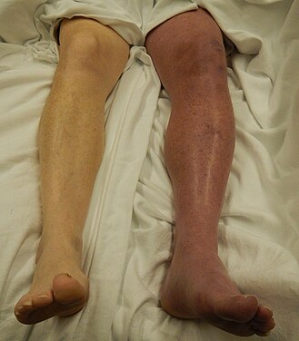
A case of phlegmasia cerulea dolens in the left leg
syndrome-%28PSS%29-in-a-young-judo-tutor-a-case-report-13256_2016_848_Fig1_HTML.jpg" decoding="async" width="286" height="252" data-file-width="358" data-file-height="314">
A venogram before catheter-directed thrombolysis against Paget–Schroetter syndrome, a rare and severe arm DVT shown here in a judo practitioner, with highly restricted blood flow shown in the vein
syndrome-%28PSS%29-in-a-young-judo-tutor-a-case-report-13256_2016_848_Fig2_HTML.jpg" decoding="async" width="246" height="252" data-file-width="358" data-file-height="366">
After treatment with catheter-directed thrombolysis, blood flow in the axillary and subclavian vein were significantly improved. Afterwards, a first rib resection provided thoracic outlet decompression to reduce the risk of recurrent DVT and the risk of sequelae from thoracic outlet compression.


The placement of an inferior vena cava filter (IVC filter) is a potential treatment option when either the standard treatment for acute DVT, anticoagulation, is absolutely contraindicated (not possible), or if someone develops a PE despite being anticoagulated. However, a 2020 NICE review found "little good evidence" for their use. A 2018 study associated IVC filter placement with a 50% reduction in PE, a 70% increase in DVT, and an 18% increase in 30 day mortality when compared to no IVC placement. As such, if someone develops a PE despite being anticoagulated, care should be given to optimize anticoagulation treatment and address other related concerns before considering the placement of a IVC filter.
A mechanical thrombectomy device can remove venous clots, although the ACCP considers it an option only when the following conditions apply: "iliofemoral DVT, symptoms for < 7 days (criterion used in the single randomized trial), good functional status, life expectancy of ≥ 1 year, and both resources and expertise are available." Anticoagulation alone is suggested over thrombectomy.
Prevention
For the prevention of blood clots in the general population, incorporating leg exercises and walking when sitting for hours at a time, having an active lifestyle, and maintaining a healthy body weight are recommended. Walking increases blood flow through the leg veins. Excess body weight is modifiable unlike most risk factors, and interventions or lifestyle modifications that help someone who is overweight or obese lose weight reduce DVT risk. Statins have been investigated for primary prevention, and the JUPITER trial, which used rosuvastatin, has provided some tentative evidence of effectiveness. Out of all the statins that have been studied, rosuvastatin appears to be the only one with the potential to reduce VTE risk. However, the number needed to treat to prevent one initial VTE is about 2000, limiting its applicability.
After VTE
Anticoagulation, which increases the risk of bleeding, is sometimes used indefinitely (lifelong treatment) in those with a high-risk for recurrence. The risk of major bleeding with long-term anticoagulation is about 3% per year, and the point where annual VTE risk is thought to warrant long-term anticoagulation is estimated to be between 3 and 9%. Usually, only when individuals exceed a 9% annual VTE risk is long-term anticoagulation a common consideration. For example, antithrombin deficiency, a strong or moderately strong risk factor, carries an annual risk of VTE of only 0.8–1.5%; as such, asymptomatic individuals with thrombophilia do not warrant long-term anticoagulation. If someone decides to stop anticoagulation after an unprovoked VTE instead of being on lifelong anticoagulation, aspirin can be used to reduce the risk of recurrence, but it is less effective at preventing VTE than anticoagulation. Statins have also been investigated for their potential to reduce recurrent VTE rates, with some studies suggesting effectiveness.
Hospital (non-surgical) patients

Acutely ill hospitalized patients are suggested to receive a parenteral anticoagulant, although the potential net benefit is uncertain. Critically ill hospitalized patients are recommended to either receive unfractionated heparin or low-molecular weight heparin instead of foregoing these medicines.
After surgery

Major orthopedic surgery—total hip replacement, total knee replacement, or hip fracture surgery—has a high risk of causing VTE. If prophylaxis is not used after these surgeries, symptomatic VTE has about a 4% chance of developing within 35 days. Following major orthopedic surgery, a blood thinner or aspirin is typically paired with intermittent pneumatic compression, which is the preferred mechanical prophylaxis over graduated compression stockings.
Options for VTE prevention in people following non-orthopedic surgery include early walking, mechanical prophylaxis, and blood thinners (low-molecular-weight heparin and low-dose-unfractionated heparin) depending upon the risk of VTE, risk of major bleeding, and person's preferences. After low-risk surgeries, early and frequent walking is the best preventive measure.
Pregnancy
The risk of VTE is increased in pregnancy by about five times because of a more hypercoagulable state, a likely adaptation against fatal postpartum hemorrhage. Additionally, pregnant women with genetic risk factors are subject to a roughly three to 30 times increased risk for VTE. Preventive treatments for pregnancy-related VTE in hypercoagulable women were suggested by the ACCP in 2012. Homozygous carriers of factor V Leiden or prothrombin G20210A with a family history of VTE were suggested for antepartum LMWH and either LMWH or a vitamin K antagonist (VKA) for the six weeks following childbirth. Those with another thrombophilia and a family history but no previous VTE were suggested for watchful waiting during pregnancy and LMWH or—for those without protein C or S deficiency—a VKA. Homozygous carriers of factor V Leiden or prothrombin G20210A with no personal or family history of VTE were suggested for watchful waiting during pregnancy and LMWH or a VKA for six weeks after childbirth. Those with another thrombophilia but no family or personal history of VTE were suggested for watchful waiting only. Warfarin, a common VKA, can cause harm to the fetus and is not used for VTE prevention during pregnancy.
Travelers

Suggestions for at-risk long-haul travelers include calf exercises, frequent walking, and aisle seating in airplanes to ease walking. Graduated compression stockings have sharply reduced the levels of asymptomatic DVT in airline passengers, but the effect on symptomatic DVT, PE, or mortality is unknown, as none of the individuals studied developed these outcomes. However, graduated compression stockings are not suggested for long-haul travelers (>4 hours) without risk factors for VTE. Likewise, neither aspirin nor anticoagulants are suggested in the general population undertaking long-haul travel. Those with significant VTE risk factors undertaking long-haul travel are suggested to use either graduated compression stockings or LMWH for VTE prevention. If neither of these two methods are feasible, then aspirin is suggested.
Prognosis
DVT is most frequently a disease of older age that occurs in the context of nursing homes, hospitals, and active cancer. It is associated with a 30-day mortality rate of about 6%, with PE being the cause of most of these deaths. Proximal DVT is frequently associated with PE, unlike distal DVT, which is rarely if ever associated with PE. Around 56% of those with proximal DVT also have PE, although a chest CT is not needed simply because of the presence of DVT. If proximal DVT is left untreated, in the following 3 months approximately half of people will experience symptomatic PE.
Another frequent complication of proximal DVT, and the most frequent chronic complication, is post-thrombotic syndrome, where individuals have chronic venous symptoms. Symptoms can include pain, itching, swelling, paresthesia, a sensation of heaviness, and in severe cases, leg ulcers. After proximal DVT, an estimated 20–50% of people develop the syndrome, with 5–10% experiencing severe symptoms. Post-thrombotic syndrome can also be a complication of distal DVT, though to a lesser extent than with proximal DVT.
Recurrence of DVT is another potential consequence. In the 10 years following an initial VTE, about 30% of people will have a recurrence. VTE recurrence in those with prior DVT is more likely to recur as DVT than PE. Cancer and unprovoked DVT are strong risk factors for recurrence. After initial proximal unprovoked DVT with and without PE, 16–17% of people will have recurrent VTE in the 2 years after they complete their course of anticoagulants. VTE recurrence is less common in distal DVT than proximal DVT. In upper extremity DVT, annual VTE recurrence is about 2–4%. After surgery, a provoked proximal DVT or PE has an annual recurrence rate of only 0.7%.
Epidemiology
About 1.5 out of 1000 adults a year have a first VTE in high-income countries, and about 5–11% of people will develop VTE in their lifetime. VTE becomes much more common