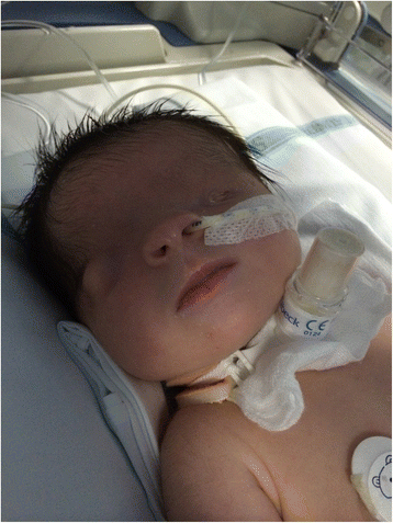
Fraser Syndrome 1

A number sign (#) is used with this entry because Fraser syndrome-1 (FRASRS1) is caused by homozygous or compound heterozygous mutation in the FRAS1 gene (607830) on chromosome 4q21.
DescriptionFraser syndrome is an autosomal recessive malformation disorder characterized by cryptophthalmos, syndactyly, and abnormalities of the respiratory and urogenital tract (summary by van Haelst et al., 2008).
Genetic Heterogeneity of Fraser Syndrome
Fraser syndrome-2 (FRASRS2) is caused by mutation in the FREM2 gene (608945) on chromosome 13q13, and Fraser syndrome-3 (FRASRS3) is caused by mutation in the GRIP1 gene (604597) on chromosome 12q14.
See Bowen syndrome (211200) for a comparable but probably distinct syndrome of multiple congenital malformations.
Clinical FeaturesIn each of 2 sibships, Fraser (1962) observed 2 sisters affected at birth by various combinations: cryptophthalmos; absent or malformed lacrimal ducts; middle and outer ear malformations; high palate; cleavage along the midplane of nares and tongue; hypertelorism; laryngeal stenosis; syndactyly; wide separation of symphysis pubis; displacement of umbilicus and nipples; primitive mesentery of small bowel; maldeveloped kidneys; fusion of labia and enlargement of clitoris; and bicornuate uterus and malformed fallopian tubes. In each sibship, 1 sister was stillborn and the other viable. Sex chromatin was positive in both surviving infants. Neither set of parents was consanguineous.
Gupta and Saxena (1962) reported cryptophthalmos in 2 offspring of consanguineous parents. In 1 case it was unilateral and death occurred at 1 month. In the other the cryptophthalmos was bilateral and was accompanied by congenital deafness, undescended testes, small penis with hypospadias, and other deformities. The older literature on cryptophthalmos with associated malformations was reviewed by Duke-Elder (1963).
Francois (1969) described affected brother and sister and gave a comprehensive review, pointing out the rather frequent examples of parental consanguinity (about 15% of cases) and of familial cases. Syndactyly is a feature of many of the cases. Francannet et al. (1990) reported instances of parental consanguinity: in 1 family, 3 children were born of a first-cousin marriage, and in a second, an affected fetus resulted from a double first-cousin marriage. An isolated case was reported by Ide and Wollschlaeger (1969). The parents were not related. The patient had syndactyly, hair extending forward over the temples, deformity of the nares and external auditory meatus, etc. Azevedo et al. (1973) reported 4 cases in 2 sibships, each with consanguineous parents. Syndactyly and other malformations were present in some.
Lurie and Cherstvoy (1984) reported 4 families with the cryptophthalmos-syndactyly syndrome. Perinatal death occurred in 9 affected members. Autopsy, performed in 6 cases, showed renal agenesis (bilateral in 3, unilateral in 3). One family had at least 4 affected sibs and 2 others had 2 affected children each. No consanguinity was established.
Burn and Marwood (1982) reported 3 sibs with Fraser syndrome presenting as bilateral renal agenesis. Only 1 of the 3 sibs had cryptophthalmos (which was unilateral). Because cryptophthalmos is not an essential part of the syndrome, Fraser syndrome is a preferable designation.
Mortimer et al. (1985) reported monozygotic twins concordant for bilateral renal agenesis and other features consistent with Fraser syndrome: syndactyly and laryngeal stenosis.
From their personal experience and a review of the literature, Koenig and Spranger (1986) identified 5 cases of the cryptophthalmos-syndactyly syndrome without cryptophthalmos. Thomas et al. (1986) also emphasized the occurrence of the cryptophthalmos syndrome without cryptophthalmos--an argument for using the eponym Fraser syndrome. In a literature review, they found 124 cases of cryptophthalmos of which 27 were isolated and 97 associated with multiple malformations. They found that isolated cryptophthalmos was sporadic in 16 cases and familial in 11. The familial cases were in 3 families with vertical transmission (123570). Some of the most characteristic malformations of the Fraser syndrome occur in areas that remain temporarily fused in utero: the eyelids, the digits, and the vagina. Since separation of the eyelids and digits involves a process of controlled necrosis, Thomas et al. (1986) speculated about a defect in programmed cell death. They also drew attention to the parallel with experimental teratogenesis by means of hypovitaminosis A in the pig and rat. Malformations reminiscent of the Fraser syndrome are produced. Thomas et al. (1986) asked whether defects in the metabolism of retinoids may play a role in pathogenesis. Meinecke (1986) also contributed to the understanding of the Fraser syndrome without cryptophthalmos.
Greenberg et al. (1986) described associated gonadal dysgenesis in an infant girl with Fraser syndrome: gonadoblastoma developed in a dysgenetic ovary.
Gattuso et al. (1987) gave a quantitative estimate of the frequency of the several clinical manifestations of Fraser syndrome on the basis of 3 new cases and 68 published cases. Craniofacial abnormalities were reported in all, cryptophthalmos in 93%, and syndactyly in 54%.
Bialer and Wilson (1988) reported a child who according to the strict criteria proposed by Thomas et al. (1986) might not be considered to have syndromal cryptophthalmos; however, the child had several malformations considered minor in that classification and was born of parents who were related in a complex way, giving the proband a coefficient of consanguinity of 0.086.
Boyd et al. (1988) presented postmortem findings in 11 cases of probable Fraser syndrome; 8 were neonatal deaths, 1 was a stillbirth, and 2 were midtrimester fetuses. Ramsing et al. (1990) presented the clinical and autopsy findings in 2 fetuses and 1 newborn infant with Fraser syndrome.
Stevens et al. (1994) observed pulmonary hyperplasia and laryngeal stenosis in 2 sibs with Fraser cryptophthalmos syndrome. Markedly enlarged echogenic lungs were demonstrable at 16 and 17 weeks of gestation. A third unrelated patient with Fraser syndrome had laryngeal stenosis, renal agenesis, and normal lung development. Reports of 3 additional cases of pulmonary hyperplasia in the Fraser syndrome were found. In all of these cases the likely mechanism for pulmonary hyperplasia was retention of fetal lung fluid by laryngeal or tracheal obstruction.
Pankau et al. (1994) described a newborn female with total syndactyly of the 3rd and 4th fingers, partial syndactyly of all other fingers, postaxial polydactyly of feet, bilateral renal agenesis, and bilateral anophthalmia. The child was born of consanguineous Turkish parents. Although the infant had normal eyelids with palpebrae and eyelashes and had histologically confirmed anophthalmia and postaxial polydactyly, Pankau et al. (1994) considered this case to be an unusual manifestation of Fraser syndrome.
Amr (1996) described unilateral cryptophthalmos with bilateral renal agenesis and syndactyly of hands and feet in a stillborn female fetus.
Andiran et al. (1999) reported a case of Fraser syndrome with anterior urethral atresia. They suggested that newborns with Fraser syndrome, especially those with umbilical discharge or severe hydroureteronephrosis, be evaluated for patency of the anterior urethra.
Slavotinek and Tifft (2002) reviewed 117 cases diagnosed as Fraser syndrome or cryptophthalmos published after the comprehensive review of Thomas et al. (1986). Their findings emphasized the clinical variability associated with Fraser syndrome and supported heterogeneity of the disorder. They also noted patterns of anomalies (for example, bicornuate uterus with imperforate anus or anal stenosis and renal malformations) that are found in other syndromes and associations without cryptophthalmos, suggesting that common modifier genes may explain some of the phenotypic variation in Fraser syndrome.
Van Haelst et al. (2007) studied the clinical features of 59 newly diagnosed individuals with Fraser syndrome from 40 families, including 25 consanguineous families. Compared to previous reviews, they found a higher frequency of abnormalities of the skull, larynx, umbilicus, urinary tract, and anus, and a lower frequency of mental retardation and cleft lip with or without cleft palate. Clinical features in probands and sibs were remarkably similar. Prenatally diagnosed patients had more manifestations giving rise to a pathologic amount of amniotic fluid, but otherwise had similar frequency of symptoms to those diagnosed postnatally.
DiagnosisKoenig and Spranger (1986) noted that eye lesions are apparently nonobligatory components of the syndrome. The diagnosis of Fraser syndrome should be entertained in patients with a combination of acrofacial and urogenital malformations with or without cryptophthalmos. Thomas et al. (1986) also emphasized the occurrence of the cryptophthalmos syndrome without cryptophthalmos and proposed a diagnostic criteria for Fraser syndrome. Major criteria consisted of cryptophthalmos, syndactyly, abnormal genitalia, and positive family history. Minor criteria were congenital malformation of the nose, ears, or larynx, cleft lip and/or palate, skeletal defects, umbilical hernia, renal agenesis, and mental retardation. Diagnosis was based on the presence of at least 2 major and 1 minor criteria, or 1 major and 4 minor criteria.
Boyd et al. (1988) suggested that prenatal diagnosis by ultrasound examination of eyes, digits, and kidneys should detect the severe form of the syndrome. Serville et al. (1989) demonstrated the feasibility of ultrasonographic diagnosis of the Fraser syndrome at 18 weeks' gestation. They suggested that the diagnosis could be made if 2 of the following signs are present: obstructive uropathy, microphthalmia, syndactyly, and oligohydramnios. Schauer et al. (1990) made the diagnosis at 18.5 weeks' gestation on the basis of sonography. Both the female fetus and the phenotypically normal father had a chromosome anomaly: inv(9)(p11q21). An earlier born infant had Fraser syndrome and the same chromosome 9 inversion.
Van Haelst et al. (2007) provided a revision of the diagnostic criteria for Fraser syndrome according to Thomas et al. (1986) through the addition of airway tract and urinary tract anomalies to the major criteria and removal of mental retardation and clefting as criteria. Major criteria included syndactyly, cryptophthalmos spectrum, urinary tract abnormalities, ambiguous genitalia, laryngeal and tracheal anomalies, and positive family history. Minor criteria included anorectal defects, dysplastic ears, skull ossification defects, umbilical abnormalities, and nasal anomalies. Cleft lip and/or palate, cardiac malformations, musculoskeletal anomalies, and mental retardation were considered uncommon. Van Haelst et al. (2007) suggested that the diagnosis of Fraser syndrome can be made if either 3 major criteria, or 2 major and 2 minor criteria, or 1 major and 3 minor criteria are present in a patient.
MappingBy autozygosity mapping, McGregor et al. (2003) located the Fraser syndrome-1 locus to chromosome 4q21.
Molecular GeneticsIn 5 families with Fraser syndrome, McGregor et al. (2003) identified 5 homozygous mutations in the FRAS1 gene (e.g., 607830.0001), which encodes a putative extracellular matrix (ECM) protein.
In a female infant with Fraser syndrome, Slavotinek et al. (2006) identified compound heterozygosity for a deletion (607830.0006) and an insertion (607830.0007) in the FRAS1 gene, inherited from her mother and her father, respectively.
Cavalcanti et al. (2007) described 2 stillborn Brazilian male sibs, born at 25 and 29 weeks' gestation, respectively. One sib appeared to have a lethal form of ablepharon-macrostomia syndrome (AMS; 200110) or an intermediate phenotype between AMS and Fraser syndrome, and the other had classic Fraser syndrome. Analysis of the FRAS1 gene revealed homozygosity for a splice site mutation (607830.0008), resulting in a severely truncated protein in both sibs and heterozygosity for the mutation in both parents. Cavalcanti et al. (2007) concluded that a phenotype resembling AMS is a rare clinical expression of Fraser syndrome, with no obvious genotype/phenotype correlation.
Animal ModelWinter (1990) speculated that Fraser syndrome is a human equivalent of the 'blebbed' phenotype in the mouse, which has been associated with mutations in at least 5 loci, including bl (Darling and Gossler, 1994). McGregor et al. (2003) screened DNA from bl/bl mice and identified a mutation in the Fras1 gene that resulted in premature protein termination. Thus, the bl mouse is a model for Fraser syndrome in humans.
Vrontou et al. (2003) showed that bl/bl homozygous embryos were devoid of Fras1 protein, consistent with the finding that Fras1 is mutated in these mice. The data suggested that perturbations in the composition of extracellular space underlying epithelia can account for the onset of the blebbed phenotype in mice and Fraser syndrome in humans.
Takamiya et al. (2004) presented data indicating that glutamate receptor-interacting protein-1 (GRIP1; 604597) is required for normal cell-matrix interactions during early embryonic development and that inactivation of Grip1 causes Fraser syndrome-like defects in mice. Kiyozumi et al. (2006) showed that Grip1-mutant 'eye blebs' (eb) mice had reduced localization of Fras1, Frem1 (608944), and Frem2 to epidermal basement membranes.
To investigate the cause of Fraser syndrome not linked to FRAS1, Jadeja et al. (2005) carried out linkage analysis in the mouse 'myelencephalic blebs' (my) strain, which shows a phenotype similar to that of Fras1-mutant mice. The authors mapped the my phenotype to the Frem2 gene and showed that a Frem2 gene trap mutation was allelic to my.
Kiyozumi et al. (2006) found that my/my mice had reduced localization of Frem2, as well as Fras1 and Frem1, to epidermal basement membranes. Frem1-knockout mice had reduced expression of Fras1 and Frem2 at the basement membrane. When coexpressed and secreted by transfected cells, these proteins formed a ternary complex, raising the possibility that their reciprocal stabilization at the basement membrane was due to complex formation. Kiyozumi et al. (2006) suggested that coordinated assembly of the 3 Fraser syndrome-associated proteins at the basement membrane is instrumental in epidermal-dermal interactions during morphogenetic processes.
HistoryGattuso et al. (1987) quoted Warkany (1971) as citing the description by Pliny the Elder in the first century A.D. of a family with 3 children born with a membrane over the eye.
Elcioglu and Berry (2000) described Fraser syndrome in a museum specimen at Guy's Hospital. The specimen had been acquired about 10 years before the description of the entity by Fraser (1962).
On the occasion of the retirement of George Fraser, Reardon (1997) provided a biographical and historical note on Fraser's contributions to clinical genetics.