Thrombotic Thrombocytopenic Purpura, Congenital

A number sign (#) is used with this entry because familial thrombotic thrombocytopenic purpura (TTP) is caused by mutation in the ADAMTS13 gene (604134), which encodes the von Willebrand factor (VWF; 613160)-cleaving protease (VWFCP).
See 235400 for a discussion of the hemolytic-uremic syndrome (HUS), which has signs and symptoms similar to those in thrombotic thrombocytopenic purpura.
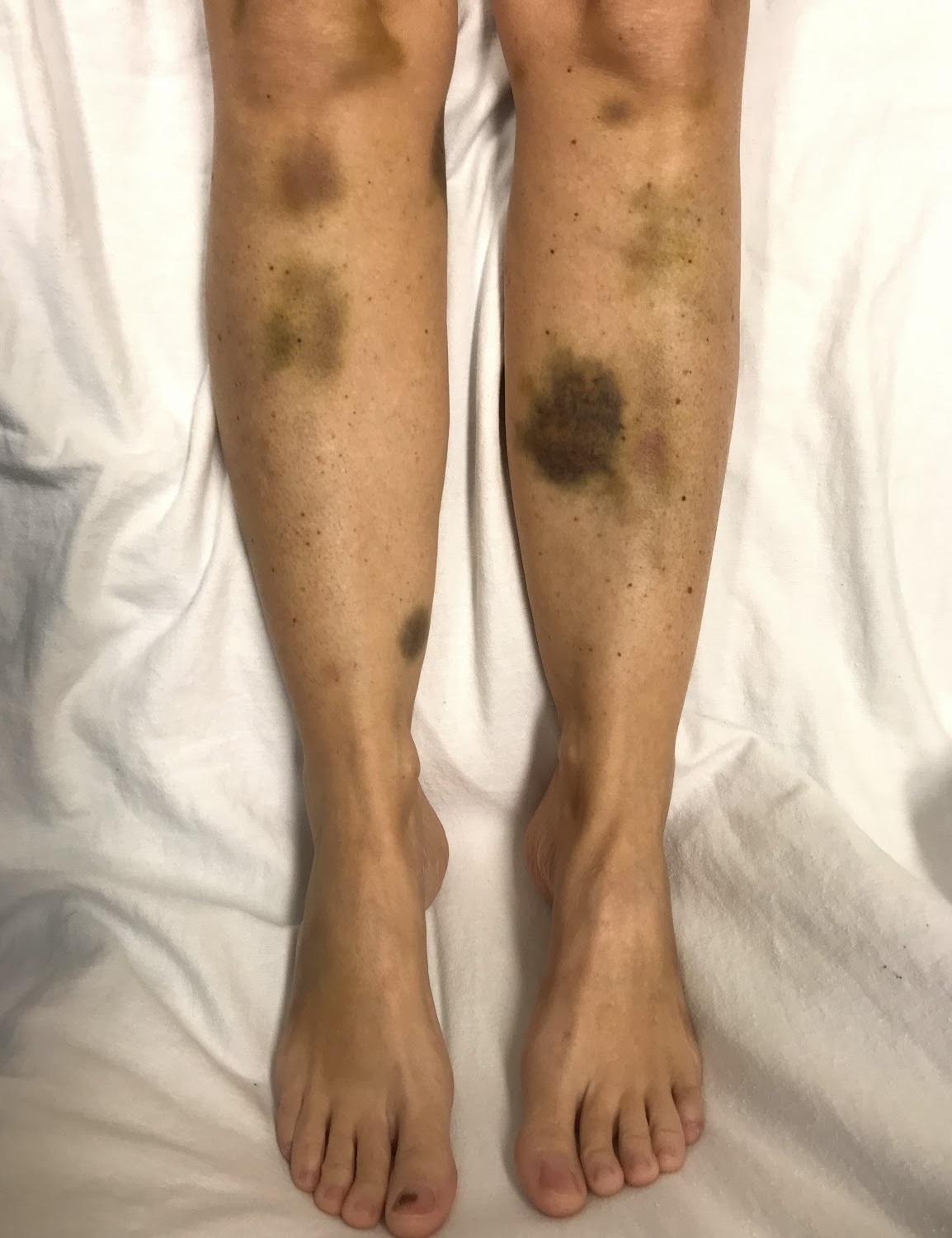
DescriptionThe classic pentad of TTP includes hemolytic anemia with fragmentation of erythrocytes, thrombocytopenia, diffuse and nonfocal neurologic findings, decreased renal function, and fever. Congenital TTP, also known as Schulman-Upshaw syndrome, is characterized by neonatal onset, response to fresh plasma infusion, and frequent relapses (Savasan et al., 2003; Kokame et al., 2002).
Acquired TTP, which is usually sporadic, usually occurs in adults and is caused by an IgG inhibitor against the von Willebrand factor-cleaving protease.
Clinical FeaturesUpshaw (1978) described a female with congenital deficiency of a factor in normal plasma that reverses microangiopathic hemolysis and thrombocytopenia, indicating a factor important to platelet and red cell survival. The proband, an only child of unrelated parents, was born with rudimentary right radius and ulna and a lobster claw deformity of the right hand. For the first 12 years of life she had 6 to 10 episodes a year of high fever, petechial rash, severe thrombocytopenia, and severe anemia. She would respond dramatically to blood transfusion, whereas adrenocorticosteroids and splenectomy were of no avail. After age 12, the attacks decreased to 3 or 4 yearly.
The same disorder may have been present in the patient of Schulman et al. (1960), an 8-year-old girl who had thrombocytopenia which responded to transfusions of blood or plasma. Deficiency of a stimulating factor that is responsible for megakaryocyte maturation and platelet production was postulated. The family history was negative. The mother's plasma induced normal platelet responses, whereas the father's resulted in submaximal responses. The patient of Schulman et al. (1960) was studied by a number of physicians because she moved from city to city. Splenectomy was of no benefit. In 1965, after a 5-month period of thrombocytopenia during which she did not receive intravenous plasma infusions, she had a complex of symptoms resembling those of glomerulonephritis, which was confirmed by renal biopsy (Abildgaard and Simone, 1967). The symptoms remitted with the reintroduction of plasma therapy. In 1973, the patient had preeclampsia during a pregnancy that resulted in a full-term normal boy. McDonald (1977) also postulated deficiency of a thrombopoietin-like substance in this patient. Plasma saved in 1975 and 1976 from this patient had normal levels of fibronectin (Goodnough et al., 1982). Rennard and Abe (1979) demonstrated deficiency of cold-insoluble globulin (fibronectin) in the patient of Upshaw (1978) but not in 4 other patients with thrombotic thrombocytopenic purpura.
Koizumi et al. (1981) described a patient who had thrombocytopenia and microangiopathic hemolytic anemia that seemed to improve with plasma administration. The plasma concentration of fibronectin was normal and intravenous administration of fibronectin was of no benefit. Shinohara et al. (1982) reported the case of a Japanese girl with similar clinical features responsive to plasma infusions. Hemolytic anemia, thrombocytopenia, distorted and fragmented circulating red cells, and megakaryocytosis of the bone marrow were present from the newborn period. They called the condition 'congenital microangiopathic hemolytic anemia' and suggested it was different from thrombotic thrombocytopenic purpura.
Of 4 affected sibs (2 male, 2 female) described by Wallace et al. (1975), the disease was fatal in 3. Kirchner et al. (1982) described this disorder in mother and daughter. The daughter's illness, characterized primarily by renal insufficiency was most compatible with adult hemolytic uremic syndrome and the mother's illness, which included neurologic findings and fever, was most compatible with thrombotic thrombocytopenic purpura. Merrill et al. (1985) reported 2 certain cases of thrombotic microangiopathy and 3 possible ones in 2 generations of a North Carolina black family. All affected members presented with acute renal failure and accelerated hypertension.
Kinoshita et al. (2001) reported 2 unrelated girls with onset of symptoms of USS at ages 4 years and 11 months, respectively. One of the girls developed a right hemiparesis caused by thrombotic occlusion of the left internal carotid artery at the age of 11 years. Both girls had received fresh frozen plasma infusion every 2 weeks.
Levy et al. (2001) studied 4 pedigrees with TTP. All patients presented at birth, except for 2 who experienced their first episode of TTP at ages 4 and 8 years; however, both of these individuals had sibs with disease onset as neonates. All patients had a chronic relapsing course and responded to plasma infusion. Activity of von Willebrand factor-cleaving protease (VWFCP) (see PATHOGENESIS) was measured in the plasma of 7 affected individuals and was found to be 2 to 7% of normal; none of the patients tested positive for inhibitors. Plasma levels of the protease in the parents of the affected individuals were 0.51 to 0.68 units/ml, consistent with a heterozygous carrier state. Levels for at-risk sibs of the patients and parents fell into a bimodal distribution, with one peak consistent with carriers and the other indistinguishable from the normal distribution.
Moake (2002) reviewed thrombotic microangiopathies. Familial TTP is associated with plasma levels of ADAMTS13 activity less than 5% of normal. The disease usually presents in infancy or childhood but sometimes is not evident until much later (Furlan and Lammle, 2001). Autoantibodies against ADAMTS13 are found in some cases of acquired idiopathic TTP. There is an association with the drug ticlopidine.
Upshaw-Schulman (USS) was originally reported as a disease complex with repeated episodes of thrombocytopenia and hemolytic anemia that quickly responded to infusions of fresh frozen plasma. Clinical signs often develop in the patients during the newborn period or early infancy. Indeed, the earliest and most frequently encountered clinical manifestation is severe hyperbilirubinemia with negative Coombs test soon after birth, which requires exchange blood transfusions. Pediatric hematologists had long been more familiar with this disease than general physicians, and a variety of alternative designations were given to the disease, such as chronic relapsing TTP, congenital microangiopathic hemolytic anemia (MAHA), and familial TTP/HUS, the last because the features of thrombotic thrombocytopenic purpura were almost indistinguishable from those of hemolytic-uremic syndrome (235400) (Matsumoto et al., 2004).
Pregnancy
Fujimura et al. (2008) reported 9 Japanese women from 6 families with genetically confirmed USS who were diagnosed with the disorder during their first pregnancy. Six of the 9 had episodes of thrombocytopenia during childhood misdiagnosed as autoimmune idiopathic thrombocytopenic purpura (AITP; 188030). Thrombocytopenia occurred during the second to third trimesters in each of their 15 pregnancies, often followed by TTP. Of 15 pregnancies, 8 babies were stillborn or died soon after birth, and the remaining 7 were all premature except 1, who was born naturally following plasma infusions to the mother that had started at 8 weeks' gestation. All women had severely deficient ADAMTS13 activity. Fujimura et al. (2008) emphasized the importance of measuring ADAMTS13 activity in the evaluation of thrombocytopenia during childhood and pregnancy.
InheritanceFurlan et al. (1997) reported 2 brothers with chronic relapsing TTP who were deficient in the VWF-cleaving protease. In addition to these brothers, Furlan et al. (1998) found complete protease deficiency in 3 sibs: 2 sisters had their first episode of TTP during pregnancy, whereas their protease-deficient brother was asymptomatic for the disorder. Three further unaffected sibs of the family (2 brothers and 1 sister) had normal activity of VWF-cleaving protease. A third family with 2 affected brothers was reported. No consanguinity was established in any of the 3 families Furlan (1999). Autosomal recessive inheritance of the disorder was suggested.
PathogenesisMoake et al. (1982) found unusually large multimers of von Willebrand factor (ULVWFMs) in the plasma of 4 patients, including the girl reported by Schulman et al. (1960), with chronic relapsing thrombotic thrombocytopenic purpura and proposed that these are the 'agglutinative' substances. These unusually large multimers are even larger than the largest multimers of VWF in normal plasma and resemble a subgroup of huge VWF forms secreted by human endothelial cells. After retrograde secretion by endothelial cells, these unusually large multimers become entangled in subepithelial fibrous components, thereby maximizing VWF-mediated adhesion of platelets to subendothelium after vascular damage. Normally, a processing activity in plasma prevents the highly adhesive, unusually large multimers from going far or staying long after being secreted into the bloodstream. Moake (1998) proposed that patients with chronic relapsing thrombotic thrombocytopenic purpura have a defect in the processing of these unusually large multimers that makes them susceptible to periodic relapses.
Furlan et al. (1996) and Tsai (1996) independently reported that a metal-containing proteolytic enzyme (metalloprotease) in normal plasma cleaves the peptide bond between tyrosine at position 842 and methionine at position 843 in monomeric subunits of VWF, thereby degrading the large multimers. This von Willebrand factor-cleaving protease was found by Furlan et al. (1997) to be deficient in 4 patients with chronic relapsing thrombotic thrombocytopenic purpura, 2 of whom were brothers. Because no inhibitor of the enzyme was detected in plasma, the deficiency was ascribed to an abnormality in the production, survival, or function of the protease.
Furlan et al. (1998) studied plasma samples from 30 patients with TTP and 23 patients with the hemolytic-uremic syndrome. Of 24 patients with nonfamilial TTP, 20 had severe and 4 had moderate protease deficiency during an acute event. An inhibitor of VWF found in 20 of the 24 patients (in all 5 plasma samples tested) was shown to be an IgG antibody. Furlan et al. (1998) found that 6 patients with familial TTP lacked VWFCP activity but had no inhibitor, whereas all 10 patients with familial hemolytic-uremic syndrome had normal protease activity. In vitro proteolytic degradation of von Willebrand factor by the protease was studied in 5 patients with familial and 7 patients with nonfamilial hemolytic-uremic syndrome and was found to function normally in all 12 patients. Furlan et al. (1998) concluded that nonfamilial TTP is due to an inhibitor of VWFCP, whereas the familial form is caused by a constitutional deficiency of the protease. Patients with the hemolytic-uremic syndrome do not have a deficiency of VWFCP or a defect in von Willebrand factor that leads to its resistance to protease.
Tsai and Lian (1998) found severe deficiency of von Willebrand factor-cleaving protease in 37 patients with acute thrombotic thrombocytopenic purpura. No deficiency was detected in 16 samples of plasma from patients in remission. Inhibitory activity against the protease was detected in 26 of 39 plasma samples obtained during the acute phase of the disease. The inhibitors were IgG antibodies.
Tati et al. (2013) demonstrated deposition of complement C3 (120700) and C5b (120900)-C9 (120940) in renal cortex of 2 TTP patients using immunofluorescence microscopy and immunohistochemical analysis, respectively. Flow cytometric analysis showed that plasma from TTP patients contained significantly higher levels of complement-coated endothelial particles than control plasma. Histamine-stimulated glomerular endothelial cells exposed to patient platelet-rich plasma or patient platelet-poor plasma combined with normal platelets induced C3 deposition, via the alternative pathway, on VWF platelet strings and on endothelial cells in an in vitro perfusion system under shear conditions. No complement was detected when cells were exposed to control plasma or to patient plasma treated with EDTA or that had been heat inactivated. Tati et al. (2013) concluded that the microvascular process induced by ADAMTS13 deficiency triggers complement activation on platelets and endothelium and may contribute to thrombotic microangiopathy.
Clinical ManagementIn the 2 patients with USS reported by Kinoshita et al. (2001), Yagi et al. (2001) studied the relationship between ULVWFMs and thrombocytopenia by analyzing platelet aggregation using a mixture of the patients' plasma and normal washed platelets under high shear stress. There was a remarkably enhanced high shear stress-induced platelet aggregation by the patients' plasma, which was almost completely normalized by administration of fresh frozen plasma. The results indicated that thrombocytopenia in USS patients is caused by a combination of the presence of ULVWFMs, platelets, and high shear stress generated in the microcirculation.
Vesely et al. (2003) stated that initial management of patients with TTP is difficult because of lack of specific diagnostic criteria, high mortality without plasma exchange treatment, and risks of plasma exchange. They performed a prospective study of ADAMTS13 activity in 142 consecutive patients, making measurements before beginning plasma exchange treatment. Severe ADAMTS13 deficiency, defined in this study as ADAMTS13 activity levels less than 5% of normal, was found in 18 (13%) of the 142 patients; it occurred only among pregnant/postpartum (2 of 10) and idiopathic (16 of 48) patients. Among the 48 patients with idiopathic TTP, the presenting features and clinical outcomes of the 16 who had severe ADAMTS13 deficiency were variable and not distinct from the 32 who did not have severe ADAMTS13 deficiency. Patients at all levels of ADAMTS13 activity apparently responded to plasma exchange treatment.
MappingLevy et al. (2001) used the plasma levels of VWF-cleaving protease as a phenotypic trait for linkage analysis. They analyzed DNA from affected individuals and other informative family members using 382 polymorphic microsatellite markers. A lod score of 5.63 at theta of 0.0 was obtained for marker D9S164 on 9q34 using a codominant model. Multipoint analysis for D9S164 and 4 flanking markers yielded a maximum lod score of 7.37 at marker D9S164.
Molecular GeneticsBy analysis of genomic DNA from patients with familial TTP, Levy et al. (2001) identified 12 mutations in the ADAMTS13 gene (604134.0001-604134.0012), accounting for 14 of the 15 disease alleles studied. Levy et al. (2001) demonstrated that deficiency of ADAMTS13 is the molecular mechanism responsible for thrombotic thrombocytopenic purpura and suggested that physiologic proteolysis of von Willebrand factor and/or other ADAMTS13 substrates is required for normal vascular homeostasis.
In 2 Japanese families with Upshaw-Schulman syndrome, characterized by congenital TTP with neonatal onset and frequent relapses, Kokame et al. (2002) reported 4 novel mutations in the ADAMTS13 gene (604134.0013-604134.0016). Activity of von Willebrand factor-cleaving protease was less than 3% of normal in all probands; VWFCP activity in heterozygous parents ranged from 30 to 60%.
In a patient with USS and severely reduced levels of VWFCP activity, Savasan et al. (2003) identified a homozygous mutation in the ADAMTS13 gene (604134.0017).
HistoryMoschcowitz (1924) described the abrupt onset of petechiae and pallor, followed rapidly by paralysis, coma, and death, in a 16-year-old girl. At autopsy, terminal arterioles and capillaries were occluded by hyaline thrombi, later determined to consist mostly of platelet without perivascular inflammation or endothelial desquamation. Moschcowitz (1924) suspected a 'powerful poison which had both agglutinative and hemolytic properties' as the cause of this disorder, now known as thrombotic thrombocytopenic purpura.