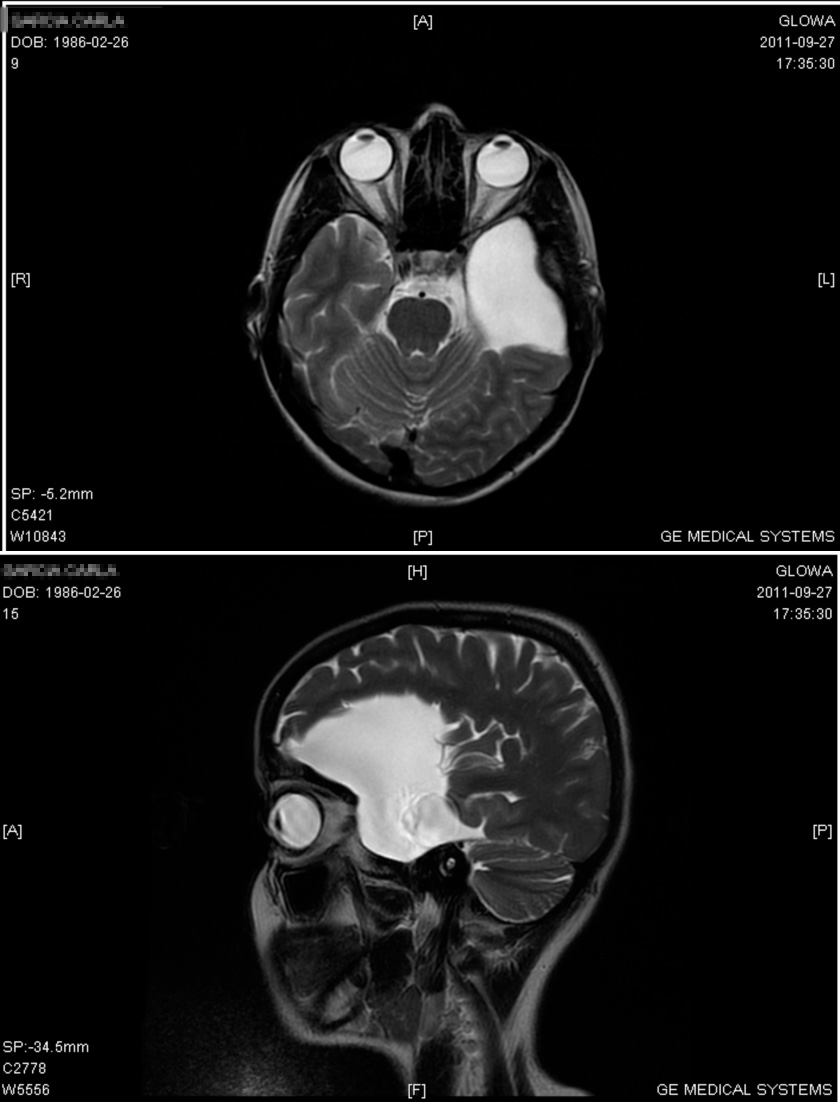
Arachnoid Cyst

A disorder with extraparenchymal cysts, intra-arachnoidal collections of fluid, the composition of which is close to that of cerebrospinal fluid. They are often asymptomatic.
Epidemiology
Studies carried out since the advent of MRI and CT suggest that the prevalence is higher than previously thought, perhaps as high as 1 per 5000. In neurosurgery case series, cysts are more commonly located in the temporo-sylvian fossae. Temporal cysts are significantly more frequent in males than in females (4:1), while cysts in other locations do not show preponderance for a specific gender. A significant predominance of left-sided temporal cysts is found in males (2:1).
Clinical description
Arachnoid cysts may evolve during postnatal life. This malformation may be primitive or may be due to a disturbed flow of cerebrospinal fluid, generated by agenesis of veins.
Etiology
Different mechanisms may explain the increase in volume of the cysts: fluid secretion by cells of the cyst wall, unidirectional valvula or fluid movements secondary to movements of vein walls.
Genetic counseling
Although most diagnosed cases are sporadic, identical arachnoid cysts have been reported in at least three sets of sibs, the parents not being related. In one family, sibs also had microcephaly and intellectual deficit. If mendelian, arachnoid cysts may be an autosomal recessive condition.