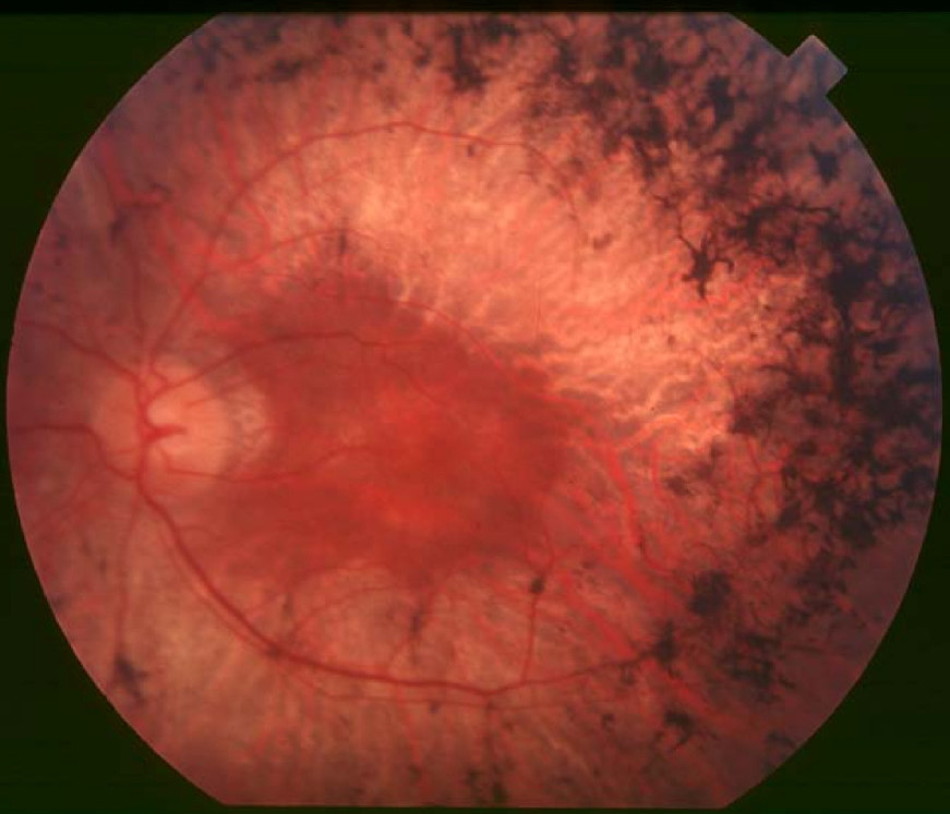
Retinitis Pigmentosa 27

A number sign (#) is used with this entry because of evidence that retinitis pigmentosa-27 (RP27) is caused by heterozygous mutation in the neural retina leucine zipper gene (NRL; 162080) on chromosome 14q11.
One family with a clinical diagnosis of clumped pigment-type retinal degeneration has been reported with compound heterozygous mutation in the NPL gene.
For a phenotypic description and a discussion of genetic heterogeneity of retinitis pigmentosa, see 268000.
Clinical FeaturesIn a large 3-generation British family (RP251) segregating autosomal dominant retinitis pigmentosa, Bessant et al. (1999) excluded previously identified autosomal dominant loci and identified a heterozygous mutation in the NRL gene (S50T; 162080.0001) in affected members. Bessant et al. (2000) identified 3 other British RP families who carried the same mutation and determined that all 4 families were descended from a common founder. In all 4 families the phenotype was fully penetrant and exhibited only limited variability. Early severe loss of rod function with preserved cone function and a very high incidence of macular edema were the characteristic features.
To further define the phenotype associated with the NRL S50T mutation, Bessant et al. (2003) performed detailed clinical, electrophysiologic, and psychophysical studies of 25 members of the original family (14 affected members, 7 unaffected, and 4 spouses). They also obtained information from clinical records for 8 individuals from the 3 families identified by Bessant et al. (2000). Bessant et al. (2003) identified 7 characteristic features of the phenotype that in combination would suggest an underlying NRL mutation. The S50T mutation causes a severe progressive retinal dystrophy affecting first the rod and subsequently the cone photoreceptors. While rod function is profoundly impaired in the first 2 decades of life, cone function remains relatively well preserved at this stage. Significant loss of cone function occurs as the disorder progresses, and in older individuals, all components of the electroretinogram are nondetectable. Patients almost invariably develop macular thickening, frequently with a mild reduction in visual acuity, between ages 15 and 30 years. As the disease progresses, a substantial loss of visual acuity is usually observed, typically in association with the development of a bull's-eye pattern of macular atrophy. Peripheral intraretinal pigment formation is sparse, even in the later stages of the disease. Distinctive peripapillary chorioretinal atrophy develops as the dystrophy progresses.
Clinical Variability
Nishiguchi et al. (2004) reported 2 sibs with a clinical diagnosis of autosomal recessive retinal degeneration of the clumped pigment type who carried compound heterozygous mutations in the NRL gene (see MOLECULAR GENETICS). Both sibs had night blindness since early childhood, consistent with a severe reduction in rod function. Color vision was normal, suggesting the presence of all cone color types; nevertheless, a comparison of central visual fields evaluated with white-on-white and blue-on-yellow light stimuli was consistent with a relatively enhanced function of short wavelength-sensitive cones in the macula. The fundi had signs of retinal degeneration (such as vascular attenuation) and clusters of large, clumped, pigment deposits in the peripheral fundus at the level of the retinal pigment epithelium.
MappingUsing linkage analysis, Bessant et al. (1999) mapped the retinitis pigmentosa phenotype in their family RP251 to chromosome 14q11, with a maximum lod score of 5.72 (theta = 0.0) at marker D14S64.
Molecular GeneticsIn a 3-generation British family with autosomal dominant retinitis pigmentosa, Bessant et al. (1999) identified a heterozygous missense mutation in the NRL gene (S50T; 162080.0001). Bessant et al. (2000) identified the S50T mutation in 3 additional families and showed that all 4 families carrying the mutation were descended from a common founder.
In 2 sibs with a clinical diagnosis of retinal degeneration of the clumped pigment type, Nishiguchi et al. (2004) identified compound heterozygous mutations in the NRL gene (224insC, 162080.0002) and (L160P, 162080.0003). Nishiguchi et al. (2004) noted that no humans with an NRL -/- genotype had previously been reported; only dominant NRL mutations that were unlikely to be null alleles had been reported. All of the published dominant NRL mutations were missense changes affecting 1 of 3 residues: ser50, pro51, or gly122. Patients with recessive NRL mutations had features resembling those caused by mutation in the NR2E3 gene (604485), the only previously known cause of enhanced S-cone syndrome (ESCS; 268100) in humans. In addition to the preservation of S-cone function, patients with recessive NR2E3 or NRL mutations have a similar pattern of intraretinal pigmentation of the fundus. This so-called clumped pigmentary retinal degeneration is found in approximately 0.5% of RP cases. Approximately half of all patients with clumped pigmentary retinal degeneration have mutations in the NR2E3 gene and are considered to have enhanced S-cone syndrome. Nishiguchi et al. (2004) concluded that mutations in NRL are a much less common cause of clumped pigmentary retinal degeneration than mutations in NR2E3.
Hernan et al. (2012) screened the NRL gene in 50 Spanish autosomal dominant RP probands and identified a heterozygous missense mutation in 1 (M96T; 162080.0004). The proband's mother and a maternal aunt were also heterozygous for the mutation; all 3 developed night blindness in the second or third decade of life, a later onset of disease than previously reported with mutations in the NRL gene. In vitro functional analysis showed that the M96T mutant increased transactivation to a lesser degree than the S50T or P51L (see Martinez-Gimeno et al., 2001) mutant proteins. In addition, the proband's sister and a cousin carried the mutation but remained asymptomatic at ages 37 and 45 years, respectively. Hernan et al. (2012) suggested that the NRL mutation might not be the sole cause of RP in this family; however, analysis of 550 retinal candidate genes by DNA capturing and massive next-generation sequencing did not reveal any other genomic variation that cosegregated with RP in the family.