Retinitis Pigmentosa 38

A number sign (#) is used with this entry because this form of retinitis pigmentosa (RP38) is caused by homozygous or compound heterozygous mutation in the MER tyrosine kinase protooncogene (MERTK; 604705) on chromosome 2q13.
DescriptionRetinitis pigmentosa (RP) describes a group of disorders with progressive degeneration of rod and cone photoreceptors in a rod-cone pattern of dysfunction. RP has a prevalence of 1 in 3,500, and is genetically and phenotypically heterogeneous (summary by Mackay et al., 2010).
For a general phenotypic description and a discussion of genetic heterogeneity of retinitis pigmentosa, see 268000.
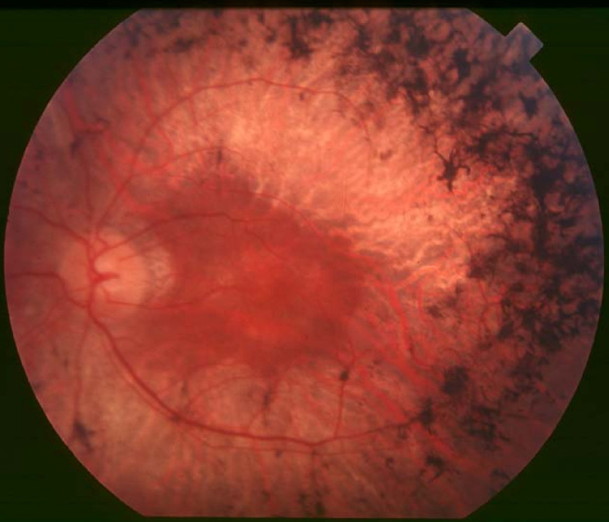
Clinical FeaturesGal et al. (2000) described 3 individuals with degenerative retinal disease who carried mutation in the MERTK gene (RP38). The first 2 patients were a 45-year-old man and a 34-year-old woman who both had onset of night blindness and poor vision in early childhood and had only a small central island of remaining vision at the time of the report. The third patient had had poor vision as a child and complained of night blindness at 12 years of age. An examination at an earlier age revealed a nondetectable rod electroretinogram and abnormally reduced visual fields. By age 17 atrophic retinal lesions were noted on ophthalmoscopic evaluation, but retinal blood vessels were not particularly attenuated.
Thompson et al. (2002) noted that the second patient described by Gal et al. (2000) had general connective tissue weakness.
Ebermann et al. (2007) clinically and genetically investigated a Moroccan family segregating both retinal dystrophy and autosomal recessive nonsyndromic hearing loss. The presence of each of these disorders in isolation in several family members suggested a partial overlap of 2 nonsyndromic autosomal recessive conditions rather than a monogenic syndrome. All affected sibs with retinal degeneration in this family exhibited severe dysfunction of both photoreceptor systems progressing with age, resulting in panretinal disease that involved the macula at an early age. The proband developed loss of visual acuity, loss of peripheral vision, and night blindness at age 11 years. Ophthalmologic examination in the sibs showed attenuated vessels in the fundus, pigmentary mottling, and drusen-like deposits in the macula. Ebermann et al. (2007) classified the retinal dystrophy as 'cone-rod dystrophy.'
Mackay et al. (2010) studied 2 brothers of Middle-Eastern origin and an unrelated Caucasian man with childhood-onset rod-cone dystrophy and early macular atrophy. The 26-year-old brother, diagnosed with rod-cone dystrophy at 16 years of age, noticed night blindness and visual field loss at 9 years, with reduced central vision at 13 years. Examination at 26 years of age revealed a visual acuity of 1.78 on the LogMAR scale and an inability to recognize any of the plates of the HRR color vision test. Funduscopy showed pale optic discs, attenuated vessels, macular atrophy, and bone spicules in the mid-periphery. His younger brother had difficulty seeing in the dark from early childhood and was diagnosed with rod-cone dystrophy at 8 years of age. His best-corrected visual acuity was 0.32 (LogMAR) bilaterally, with a low myopic refractive error, mild generalized dyschromatopsia, and visual fields that were reduced to 20 to 30 degrees bilaterally. Funduscopy revealed 'bull's eye' macular atrophy, with bone spicules and granular appearance of the retinal pigment epithelium (RPE) in the mid-periphery. Optical coherence tomography (OCT) in both sibs revealed thinning of the photoreceptor layer and multiple high-reflectance bodies below the outer limiting membrane. The unrelated Caucasian man, who noticed nyctalopia at 12 years of age and developed photophobia soon after, was diagnosed with rod-cone dystrophy at age 14. He had a strong family history of red-green colorblindness, affecting a maternal uncle and grandfather and 2 cousins, and also of deafness (mother, maternal uncle, and grandmother). At 22 years of age, he had visual acuities of 0.6 (LogMAR) on the right and 1.0 on the left, with very poor color vision. His hearing was normal, and visual fields were reported to be full at age 19. Funduscopy showed focal atrophy in the central macula and attenuated vessels but very little intraretinal pigment. OCT showed thinning of the photoreceptor layer and high reflectance bodies.
Ksantini et al. (2012) studied a consanguineous Moroccan family in which 3 sibs had the rod-cone type of retinitis pigmentosa. Salient features included night blindness starting in early infancy, dot-like whitish deposits in the fovea and macula corresponding to autofluorescent dots in the youngest patients, decreased visual acuity, and cone responses higher than rod responses on electroretinography.
Molecular GeneticsIn 2 patients with retinitis pigmentosa, Gal et al. (2000) identified homozygous mutations in the MERTK gene. The first patient, with a 5-bp deletion (604705.0001), came from a consanguineous family. The second patient, carrying a splice site mutation (604705.0002), had paternal isodisomy for chromosome 2. In a third patient a nonsense mutation was found in heterozygosity (R651X; 604705.0003); a second mutation was not found in this individual by direct sequencing of exons 1 through 19.
In 5 Moroccan sibs with retinal dystrophy, Ebermann et al. (2007) detected homozygosity for a novel splice site mutation in the MERTK gene (604705.0004). Two of these sibs were also affected with congenital progressive hearing loss (DFNB59; 610220) and carried a homozygous mutation in the pejvakin gene (610219.0003). Ebermann et al. (2007) stated that, in contrast to typical RP, all affected sibs in this family exhibited an ocular phenotype corresponding to cone-rod disease (CORD), with severe dysfunction of both photoreceptor systems that progressed with age and resulted in panretinal disease involving the macula at an early stage.
In a consanguineous Middle Eastern family segregating autosomal recessive rod-cone dystrophy, Mackay et al. (2010) performed a genomewide scan and identified 3 shared regions of homozygosity, 2 on chromosome 2 and 1 on chromosome 10. Analysis of 3 candidate genes, RGR (600342), PCDH21 (CDHR1; 609502), and MERTK, revealed no variants that segregated with disease in RGR or CDHR1, but a deletion encompassing exon 8 of the MERTK gene (604705.0005) was identified in homozygosity in the affected sibs and in heterozygosity in their unaffected parents and sibs. Screening of 100 UK probands with autosomal recessive retinal dystrophies and 100 Saudi Arabian probands with RP revealed a second Saudi Arabian family with the same exon 8 deletion. Subsequent screening of 292 probands with either Leber congenital amaurosis (LCA; see 204000) or childhood-onset retinal dystrophy identified a single 22-year-old Caucasian man who carried a known MERTK nonsense mutation, R651X; direct sequencing confirmed the R651X variant and also revealed a splice site mutation in intron 1 (604705.0006). The second Middle Eastern family was not available for examination, but review of clinical records showed that 5 affected individuals had childhood-onset RP, with visual acuity ranging from 20/50 in the first decade to hand movements only by the third decade; macular atrophy was present from the first decade and extensive peripheral atrophy and pigmentation. Mackay et al. (2010) concluded that the phenotype associated with MERTK mutations is one of childhood-onset rod-cone dystrophy and early macular atrophy, with a distinctive OCT appearance showing evidence of debris beneath the sensory retina.
In a study involving the genetically isolated Faroe Islands population, Ostergaard et al. (2011) estimated the prevalence of RP to be 1 in 1,900. SNP analysis in 21 RP patients revealed a homozygous region on chromosome 2q encompassing the MERTK gene that was common to patients in 4 families, and a 91-kb deletion in MERTK (604705.0007) was identified in 7 (30%) of the 21 patients. The 6 homozygous patients presented with onset of disease in the first decade of life followed by rapid deterioration of both rod and cone photoreceptor function, and early macular involvement was present, as seen in previously reported patients with MERTK mutations. The deletion was present in heterozygosity in 3 of 94 anonymous Faroese controls, corresponding to a carrier frequency of approximately 3%. Ostergaard et al. (2011) concluded that the 91-kb deletion represents a founder mutation in the Faroe Islands, responsible for about 30% of RP, and that mutations in the MERTK and CDHR1 genes together account for more than half of RP cases in that population.
In a consanguineous Moroccan family with rod-cone dystrophy mapping to chromosome 2, Ksantini et al. (2012) identified homozygosity for a nonsense mutation in the MERTK gene (R775X; 604705.0008). They reviewed the phenotype of 22 reported patients with MERTK mutations, noting that it appears to be a rather severe retinitis pigmentosa of the rod-cone dystrophy type, with onset in the first decade in most cases and decrease of visual acuity after 2 years of age. Discrete dot-like autofluorescent deposits are present in the early stages, and represent a hallmark of MERTK-specific retinal dystrophy.