Leber Congenital Amaurosis 3

A number sign (#) is used with this entry because Leber congenital amaurosis-3 (LCA3) and a form of juvenile retinitis pigmentosa are caused by homozygous or compound heterozygous mutation in the SPATA7 gene (609868) on chromosome 14q31.
DescriptionAutosomal recessive childhood-onset severe retinal dystrophy is a heterogeneous group of disorders affecting rod and cone photoreceptors simultaneously. The most severe cases are termed Leber congenital amaurosis, whereas the less aggressive forms are usually considered juvenile retinitis pigmentosa (Gu et al., 1997).
Mackay et al. (2011) concluded that SPATA7 retinopathy is an infantile-onset severe cone-rod dystrophy with early extensive peripheral retinal atrophy but with variable foveal involvement.
For a general phenotypic description and a discussion of genetic heterogeneity of Leber congenital amaurosis, see LCA1 (204000); for retinitis pigmentosa, see 268000.
Clinical FeaturesStockton et al. (1998) reported a large consanguineous Saudi Arabian kindred segregating Leber congenital amaurosis. In addition to poor visual acuity (typically less than 5/200), affected individuals manifested moderate midfacial hypoplasia with enophthalmos, complex vertical, horizontal, and rotatory nystagmus present from early life, moderate hypermetropic refractive errors with astigmatism, and various presentations of esotropia and exotropia. At least 4 individuals had hospital-based electroretinograms which were nonrecordable in both photopic and scotopic components in the first 2 years of life.
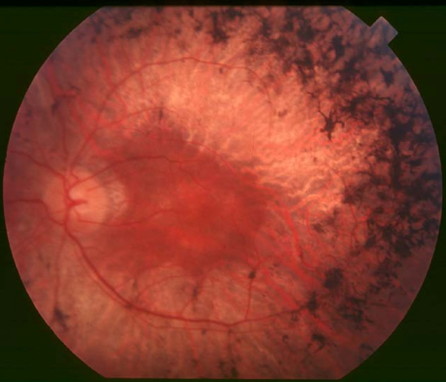
Li et al. (2009) reported a Saudi Arabian family segregating LCA. All 3 affected individuals had poor vision, nonrecordable ERGs, hypermetropic astigmatism, and infantile nystagmus. Two also showed neuroretinal atrophy and markedly attenuated vessels of the fundi.
Mackay et al. (2011) reported affected members of 5 unrelated families with LCA3. The retina had widespread retinal pigment epithelial atrophy, with minimal pigment migration into the neurosensory retina. Fundus autofluorescence imaging showed a parafoveal annulus of increased autofluorscence. High-definition optical coherence tomography showed preservation of the inner/outer segment junction at the fovea.
MappingStockton et al. (1998) used a DNA pooling strategy comparing the genotypes of affected to unaffected control pools in a genomewide search for identity by descent in a consanguineous Saudi Arabian LCA family. A shift to homozygosity was observed in the affected DNA pool compared with the control pool at linked markers D14S606 and D14S610. Testing of markers closely linked to and flanking these loci confirmed linkage with a maximum lod score of 13.29 at theta = 0.0. From these data, the LCA locus, designated LCA3, was assigned to 14q24. This locus and the previously identified loci for LCA were excluded in other Saudi Arabian pedigrees, indicating yet further genetic heterogeneity in LCA.
Using short tandem repeat markers in a Saudi Arabian family segregating LCA, Li et al. (2009) found linkage of the disorder to the LCA3 locus at 14q24 (lod score of 3.0). All 3 affected members were homozygous between markers D14S61 and D14S1066, a 13.4-Mb interval, whereas unaffected family members were heterozygous or wildtype. Li et al. (2009) noted that the haplotype in their family was different from that in the family reported by Stockton et al. (1998).
By whole-genome SNP analysis and fine mapping of the distal boundary of the LCA3 locus in 1 affected subject from each of the 2 families with LCA previously studied by Stockton et al. (1998) and Li et al. (2009), Wang et al. (2009) refined the critical region of homozygosity to 3.8 Mb between markers D14S1022 and D14S1005 on chromosome 14q31.3. The critical region contained 9 candidate genes.
Molecular GeneticsIn 2 unrelated Saudi Arabian families (KKESH-019 and KKESH-060) previously reported by Stockton et al. (1998) and Li et al. (2009), respectively, Wang et al. (2009) sequenced 9 candidate genes and identified a homozygous mutation in the SPATA7 gene (R108X; 609868.0001) in family KKESH-060 that segregated with the disease and was not found in 50 Saudi Arabian and 100 European samples. Mutation analysis in additional patients revealed homozygosity for the same R108X mutation in a Dutch LCA patient as well as a frameshift mutation in another LCA patient of Middle Eastern origin (609868.0002); homozygosity for 2 different nonsense and frameshift mutations in the SPATA7 gene was also identified in 2 patients with juvenile retinitis pigmentosa (609868.0003 and 609868.0004, respectively). Wang et al. (2009) noted that consistent with the observation that LCA has a more severe clinical phenotype than juvenile RP, the nonsense mutations associated with LCA are located in the middle of the SPATA7 coding region, whereas those associated with juvenile RP are located in the last 2 exons of SPATA7.
Mackay et al. (2011) screened all coding exons in the SPATA7 gene in 141 patients diagnosed with LCA or early childhood-onset severe retinal dystrophy and identified 4 disease-causing mutations in 5 families. They concluded that mutations in SPATA7 are a rare cause of childhood retinal dystrophy, accounting for 1.7% of disease in their cohort. Four consanguineous families with LCA, 3 of Pakistani and 1 of Bangladeshi origin, had a homozygous mutation in exon 5 (609868.0007) or exon 8 (609868.0002). In a nonconsanguineous British Caucasian family, 2 brothers had compound heterozygous mutations, one in exon 5 (609868.0005) and the other in exon 12 (609868.0006). One of the brothers had clinical features consistent with LCA, having severe visual loss from early infancy, pendular nystagmus, and sluggish pupillary responses. His brother had a milder phenotype with onset of nystagmus at 8 weeks of age. He was able to fix and follow at this age. When older, he was noted to have severe nyctalopia and constricted visual fields. These symptoms deteriorated significantly from 14 years of age.
NomenclatureLeber congenital amaurosis-13 (612712), which is caused by mutation in the RDH12 gene (608830) on chromosome 14q23.3, was originally thought to be the same as LCA3. However, affected members of the Saudi Arabian family reported by Stockton et al. (1998) do not have mutations in the RDH12 gene.