Trisomy 18

Trisomy 18 is a chromosomal abnormality associated with the presence of an extra chromosome 18 and characterized by growth delay, dolichocephaly, a characteristic facies, limb anomalies and visceral malformations.
Epidemiology
Incidence is estimated at between 1/6000 and 1/8000 births. In utero death occurs in more than 95% of fetuses with this chromosome anomaly. For unknown reasons, the rate of survival is higher in females than in males, leading to a female predominance among live-born trisomy 18 infants.
Clinical description
Hypotonia, hyporeactivity and feeding problems (poor suction) are present in the first weeks of life and are followed by a progression to hypertonia with infants showing an apparent lack of awareness of their surroundings. Common features are intrauterine and postnatal growth delay, an emaciated appearance with hypotrophy, microcephaly with a narrow skull and dolichocephaly, microretrognathia, hypertelorism, and poorly modeled and angular ears. Foot anomalies include pes equinovarus and/or rocker-bottom feet and the fingers overlap (the fifth and second fingers with the fourth and third). Malformations are common with involvement of the eyes (microphthalmia, coloboma), heart (over 90% of cases) digestive tract (esophageal atresia, anorectal malformations), kidneys and urinary tract (hydronephrosis, uni- or bilateral agenesis). Less frequently, cleft/lip palate, arthrogryposis, radial aplasia, spina bifida and anencephaly, holoprosencephaly and omphalocele are observed.
Etiology
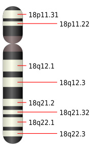
The majority of cases are associated with free trisomy 18. Mosaic trisomy 18 has been detected in a few patients presenting with a clinical picture that varies from classical trisomy 18 to a normal phenotype depending on the number of trisomic cells present in the tissues. The trisomy 18 phenotype appears to be associated with the presence of three copies of the 18q11-q12 interval.
Antenatal diagnosis
Trisomy 18 may be suspected during pregnancy from ultrasound findings (growth retardation, malformations, multiple choroid plexus cysts) and can be confirmed by karyotype analysis of the fetus. Serum markers (used for the diagnosis of trisomy 21) may also be abnormal.
Genetic counseling
The risk of recurrence of trisomy (21, 13 or 18) in families of an index case with trisomy 18 is around 1%. However, in families in which trisomy 18 is caused by translocation, the recurrence risk is higher if one of the parents is a carrier of a balanced translocation.
Management and treatment
Management is supportive only.
Prognosis
Surgical treatment of the malformations does little to improve the poor prognosis associated with this syndrome: 90% of infants die within the first year of life from cardiac, renal or neurological complications, or from repeated infections. Prolonged survival (in some cases into adulthood) has been reported, mainly in cases involving mosaic or partial trisomy (resulting from translocation). The majority of non-mosaic patients develop only limited autonomy (absence of speech and ambulation). The growth retardation is significant.