Melanoma

Melanoma, also known as malignant melanoma, is a type of skin cancer that develops from the pigment-producing cells known as melanocytes. Melanomas typically occur in the skin but may rarely occur in the mouth, intestines or eye (uveal melanoma). In women, they most commonly occur on the legs, while in men they most commonly occur on the back. About 25% of melanomas develop from moles. Changes in a mole that can indicate melanoma include an increase in size, irregular edges, change in color, itchiness or skin breakdown.
The primary cause of melanoma is ultraviolet light (UV) exposure in those with low levels of the skin pigment melanin. The UV light may be from the sun or other sources, such as tanning devices. Those with many moles, a history of affected family members and poor immune function are at greater risk. A number of rare genetic conditions such as xeroderma pigmentosum also increase the risk. Diagnosis is by biopsy and analysis of any skin lesion that has signs of being potentially cancerous.
Using sunscreen and avoiding UV light may prevent melanoma. Treatment is typically removal by surgery. In those with slightly larger cancers, nearby lymph nodes may be tested for spread (metastasis). Most people are cured if spread has not occurred. For those in whom melanoma has spread, immunotherapy, biologic therapy, radiation therapy or chemotherapy may improve survival. With treatment, the five-year survival rates in the United States are 99% among those with localized disease, 65% when the disease has spread to lymph nodes and 25% among those with distant spread. The likelihood that melanoma will reoccur or spread depends on its thickness, how fast the cells are dividing and whether or not the overlying skin has broken down.
Melanoma is the most dangerous type of skin cancer. Globally, in 2012, it newly occurred in 232,000 people. In 2015, there were 3.1 million people with active disease, which resulted in 59,800 deaths. Australia and New Zealand have the highest rates of melanoma in the world. There are also high rates in Northern Europe and North America, while it is less common in Asia, Africa and Latin America. In the United States melanoma occurs about 1.6 times more often in men than women. Melanoma has become more common since the 1960s in areas mostly populated by people of European descent.
Signs and symptoms
Early signs of melanoma are changes to the shape or color of existing moles or, in the case of nodular melanoma, the appearance of a new lump anywhere on the skin. At later stages, the mole may itch, ulcerate or bleed. Early signs of melanoma are summarized by the mnemonic "ABCDEF":
- Asymmetry
- Borders (irregular with edges and corners)
- Colour (variegated)
- Diameter (greater than 6 mm (0.24 in), about the size of a pencil eraser)
- Evolving over time
This classification does not apply to nodular melanoma, which has its own classifications:
- Elevated above the skin surface
- Firm to the touch
- Growing
Metastatic melanoma may cause nonspecific paraneoplastic symptoms, including loss of appetite, nausea, vomiting and fatigue. Metastasis (spread) of early melanoma is possible, but relatively rare: less than a fifth of melanomas diagnosed early become metastatic. Brain metastases are particularly common in patients with metastatic melanoma. It can also spread to the liver, bones, abdomen or distant lymph nodes.
Cause
Melanomas are usually caused by DNA damage resulting from exposure to ultraviolet light from the sun. Genetics also plays a role. Melanoma can also occur in skin areas with little sun exposure (i.e. mouth, soles of feet, palms of hands, genital areas). People with dysplastic nevus syndrome, also known as familial atypical multiple mole melanoma (FAMMM), are at increased risk for the development of melanoma.
Having more than fifty moles indicates an increased risk melanoma might arise. A weakened immune system makes it easier for cancer to arise due to the body's weakened ability to fight cancer cells.
UV radiation
The ultraviolet radiation from tanning beds increases the risk of melanoma. The International Agency for Research on Cancer finds that tanning beds are "carcinogenic to humans" and that people who begin using tanning devices before the age of thirty years are 75% more likely to develop melanoma.
Those who work in airplanes also appear to have an increased risk, believed to be due to greater exposure to UV.
Ultraviolet UVB light (wavelengths between 315 and 280 nm) from the sun is absorbed by skin cell DNA and results in a type of direct DNA damage called cyclobutane pyrimidine dimers (CPDs). Thymine-thymine, cytosine-cytosine or cytosine-thymine dimers are formed by the joining of two adjacent pyrimidine bases within a DNA strand. Somewhat similarly to UVB, UVA light (longer wavelengths between 400 and 315 nm) from the sun or from tanning beds can also be directly absorbed by skin DNA (at about 100 to 1000 fold lower efficiency than UVB is absorbed).
Exposure to ultraviolet radiation (UVA and UVB) is one of the major contributors to the development of melanoma. Occasional extreme sun exposure (resulting in "sunburn") is causally related to melanoma. Melanoma is most common on the back in men and on legs in women (areas of intermittent sun exposure). The risk appears to be strongly influenced by socioeconomic conditions rather than indoor versus outdoor occupations; it is more common in professional and administrative workers than unskilled workers. Other factors are mutations in or total loss of tumor suppressor genes. Use of sunbeds (with deeply penetrating UVA rays) has been linked to the development of skin cancers, including melanoma.
Possible significant elements in determining risk include the intensity and duration of sun exposure, the age at which sun exposure occurs, and the degree of skin pigmentation. Melanoma rates tend to be highest in countries settled by migrants from northern Europe that have a large amount of direct, intense sunlight that the skin of the settlers is not adapted to, most notably Australia. Exposure during childhood is a more important risk factor than exposure in adulthood. This is seen in migration studies in Australia.
Having multiple severe sunburns increases the likelihood that future sunburns develop into melanoma due to cumulative damage. The sun and tanning beds are the main sources of UV radiation that increase the risk for melanoma and living close to the equator increases exposure to UV radiation.
Genetics
A number of rare mutations, which often run in families, greatly increase melanoma susceptibility. Several genes increase risks. Some rare genes have a relatively high risk of causing melanoma; some more common genes, such as a gene called MC1R that causes red hair, have a relatively lower elevated risk. Genetic testing can be used to search for the mutations.
One class of mutations affects the gene CDKN2A. An alternative reading frame mutation in this gene leads to the destabilization of p53, a transcription factor involved in apoptosis and in fifty percent of human cancers. Another mutation in the same gene results in a nonfunctional inhibitor of CDK4, a cyclin-dependent kinase that promotes cell division. Mutations that cause the skin condition xeroderma pigmentosum (XP) also increase melanoma susceptibility. Scattered throughout the genome, these mutations reduce a cell's ability to repair DNA. Both CDKN2A and XP mutations are highly penetrant (the chances of a carrier to express the phenotype is high).
Familial melanoma (FAMMM) is genetically heterogeneous, and loci for familial melanoma appear on the chromosome arms 1p, 9p and 12q. Multiple genetic events have been related to melanoma's pathogenesis (disease development). The multiple tumor suppressor 1 (CDKN2A/MTS1) gene encodes p16INK4a – a low-molecular weight protein inhibitor of cyclin-dependent protein kinases (CDKs) – which has been localised to the p21 region of human chromosome 9. FAMMM is typically characterized by having 50 or more combined moles in addition to a family history of melanoma. It is transmitted autosomal dominantly and mostly associated with the CDKN2A mutations. People who have CDKN2A mutation associated FAMMM have a 38 fold increased risk of pancreatic cancer.
Other mutations confer lower risk, but are more common in the population. People with mutations in the MC1R gene are two to four times more likely to develop melanoma than those with two wild-type (typical unaffected type) copies. MC1R mutations are very common; and all red-haired people have a mutated copy. Mutation of the MDM2 SNP309 gene is associated with increased risks for younger women.
Fair- and red-haired people, persons with multiple atypical nevi or dysplastic nevi and persons born with giant congenital melanocytic nevi are at increased risk.
A family history of melanoma greatly increases a person's risk because mutations in several genes have been found in melanoma-prone families. People with a history of one melanoma are at increased risk of developing a second primary tumor.
Fair skin is the result of having less melanin in the skin, which means there is less protection from UV radiation. A family history could indicate a genetic predisposition to melanoma.
Pathophysiology


The earliest stage of melanoma starts when melanocytes begin out-of-control growth. Melanocytes are found between the outer layer of the skin (the epidermis) and the next layer (the dermis). This early stage of the disease is called the radial growth phase, when the tumor is less than 1 mm thick. Because the cancer cells have not yet reached the blood vessels deeper in the skin, it is very unlikely that this early-stage melanoma will spread to other parts of the body. If the melanoma is detected at this stage, then it can usually be completely removed with surgery.
When the tumor cells start to move in a different direction – vertically up into the epidermis and into the papillary dermis – cell behaviour changes dramatically.
The next step in the evolution is the invasive radial growth phase, in which individual cells start to acquire invasive potential. From this point on the melanoma is capable of spreading. The Breslow's depth of the lesion is usually less than 1 mm (0.04 in), while the Clark level is usually 2.
The vertical growth phase (VGP) following is the invasive melanoma. The tumor becomes able to grow into the surrounding tissue and can spread around the body through blood or lymph vessels. The tumor thickness is usually more than 1 mm (0.04 in), and the tumor involves the deeper parts of the dermis.
The host elicits an immunological reaction against the tumor during the VGP, which is judged by the presence and activity of the tumor infiltrating lymphocytes (TILs). These cells sometimes completely destroy the primary tumor; this is called regression, which is the latest stage of development. In certain cases, the primary tumor is completely destroyed and only the metastatic tumor is discovered. About 40% of human melanomas contain activating mutations affecting the structure of the B-Raf protein, resulting in constitutive signaling through the Raf to MAP kinase pathway.
An insult common to most cancers is damage to DNA. UVA light mainly causes thymine dimers. UVA also produces reactive oxygen species and these inflict other DNA damage, primarily single-strand breaks, oxidized pyrimidines and the oxidized purine 8-oxoguanine (a mutagenic DNA change) at 1/10th, 1/10th and 1/3rd the frequencies of UVA-induced thymine dimers, respectively.
If unrepaired, CPD photoproducts can lead to mutations by inaccurate translesion synthesis during DNA replication or repair. The most frequent mutations due to inaccurate synthesis past CPDs are cytosine to thymine (C>T) or CC>TT transition mutations. These are commonly referred to as UV fingerprint mutations, as they are the most specific mutation caused by UV, being frequently found in sun-exposed skin but rarely found in internal organs. Errors in DNA repair of UV photoproducts, or inaccurate synthesis past these photoproducts, can also lead to deletions, insertions and chromosomal translocations.
The entire genomes of 25 melanomas were sequenced. On average, about 80,000 mutated bases (mostly C>T transitions) and about 100 structural rearrangements were found per melanoma genome. This is much higher than the approximately 70 mutations across generations (parent to child). Among the 25 melanomas, about 6,000 protein-coding genes had missense, nonsense, or splice site mutations. The transcriptomes of over 100 melanomas has also been sequenced and analyzed. Almost 70% of all human protein coding genes are expressed in melanoma. Most of these genes are also expressed in other normal and cancer tissues, with some 200 genes showing a more specific expression pattern in melanoma compared to other forms of cancer. Examples of melanoma specific genes are tyrosinase, MLANA, and PMEL.
UV radiation causes damage to the DNA of cells, typically thymine dimerization, which when unrepaired can create mutations in the cell's genes. This strong mutagenic factor makes cutaneous melanoma the tumor type with the highest number of mutations. When the cell divides, these mutations are propagated to new generations of cells. If the mutations occur in protooncogenes or tumor suppressor genes, the rate of mitosis in the mutation-bearing cells can become uncontrolled, leading to the formation of a tumor. Data from patients suggest that aberrant levels of activating transcription factor in the nucleus of melanoma cells are associated with increased metastatic activity of melanoma cells; studies from mice on skin cancer tend to confirm a role for activating transcription factor-2 in cancer progression.
Cancer stem cells may also be involved.
Gene mutations
Large scale studies such as The Cancer Genome Atlas have characterized recurrent somatic alterations likely driving initiation and development of cutaneous melanoma.
The most frequent mutation occurs in the 600th codon of BRAF (50% of cases). BRAF is normally involved in cell growth and this specific mutation renders the protein constitutively active and independent of normal physiological regulation, thus fostering tumor growth. RAS genes (NRAS, HRAS and KRAS) are also recurrently mutated (30% of TCGA cases) and mutations in the 61st or 12th codons trigger oncogenic activity. Loss-of-function mutations often affect tumor suppressor genes such as NF1, TP53 and CDKN2A. Other oncogenic alterations include fusions involving various kinases such as BRAF, RAF1, ALK, RET, ROS1, NTRK1., NTRK3 and MET BRAF, RAS, NF1 mutations and kinase fusions are remarkably mutually-exclusive, as they occur in different subsets of patients. Assessment of mutation status can therefore improve patient stratification and inform targeted therapy with specific inhibitors.
In some cases (3-7%) mutated versions of BRAF and NRAS undergo copy number amplification.
Diagnosis


Looking at the area in question is the most common method of suspecting a melanoma. Moles that are irregular in color or shape are typically treated as candidates. To detect melanomas (and increase survival rates), it is recommended to learn to recognize them (see "ABCDE" mnemonic), to regularly examine moles for changes (shape, size, color, itching or bleeding) and to consult a qualified physician when a candidate appears.
However, many melanomas present as lesions smaller than 6 mm in diameter; and all melanomas are malignant when they first appear as a small dot. Physicians typically examine all moles, including those less than 6 mm in diameter. Seborrheic keratosis may meet some or all of the ABCD criteria, and can lead to false alarms. Doctors can generally distinguish seborrheic keratosis from melanoma upon examination, or with dermatoscopy.
Some advocate replacing enlarging with evolution. Certainly moles that change and evolve will be a concern. Alternatively, some practitioners prefer elevation. Elevation can help identify a melanoma, but lack of elevation does not mean that the lesion is not a melanoma. Most melanomas in the US are detected before they become elevated. By the time elevation is visible, they may have progressed to the more dangerous invasive stage.
In person inspection of suspicious skin lesions is more accurate than visual inspection of images of suspicious skin lesions. When used by trained specialists, dermoscopy is more helpful to identify malignant lesions than use of the naked eye alone. Reflectance confocal microscopy may have better sensitivity and specificity than dermoscopy in diagnosing cutaneous melanoma but more studies are needed to confirm this result.

Melanoma in skin biopsy with H&E stain – this case may represent superficial spreading melanoma.

Lymph node with almost complete replacement by metastatic melanoma. The brown pigment is focal deposition of melanin.

A dermatoscope


Ugly duckling
One method is the "ugly duckling sign". Correlation of common lesion characteristics is made. Lesions that deviate from the common characteristics are labeled an "Ugly Duckling", and a further professional exam is required. The "Little Red Riding Hood" sign suggests that individuals with fair skin and light-colored hair might have difficult-to-diagnose amelanotic melanomas. Extra care is required when examining such individuals, as they might have multiple melanomas and severely dysplastic nevi. A dermatoscope must be used to detect "ugly ducklings", as many melanomas in these individuals resemble non-melanomas or are considered to be "wolves in sheep's clothing". These fair-skinned individuals often have lightly pigmented or amelanotic melanomas that do not present easy-to-observe color changes and variations. Their borders are often indistinct, complicating visual identification without a dermatoscope.
Amelanotic melanomas and melanomas arising in fair-skinned individuals are very difficult to detect, as they fail to show many of the characteristics in the ABCD rule, break the "Ugly Duckling" sign and are hard to distinguish from acne scarring, insect bites, dermatofibromas, or lentigines.
Biopsy
Following a visual examination and a dermatoscopic exam, or in vivo diagnostic tools such as a confocal microscope, the doctor may biopsy the suspicious mole. A skin biopsy performed under local anesthesia is often required to assist in making or confirming the diagnosis and in defining severity. Elliptical excisional biopsies may remove the tumor, followed by histological analysis and Breslow scoring. Incisional biopsies such as punch biopsies are usually contraindicated in suspected melanomas, because of the possibility of sampling error or local implantation causing misestimation of tumour thickness. However, fears that such biopsies may increase the risk of metastatic disease seem unfounded.
Total body photography, which involves photographic documentation of as much body surface as possible, is often used during follow-up for high-risk patients. The technique has been reported to enable early detection and provides a cost-effective approach (with any digital camera), but its efficacy has been questioned due to its inability to detect macroscopic changes. The diagnosis method should be used in conjunction with (and not as a replacement for) dermoscopic imaging, with a combination of both methods appearing to give extremely high rates of detection.
Histopathologic types

Melanoma is a type of neuroectodermal neoplasm. There are four main types of melanoma:
| Type | Features | Incidence | Photograph | Micrograph |
|---|---|---|---|---|
| Superficial spreading melanoma | Melanoma cells with nest formation along the dermo-epidermal junction. | 70% | 
|

|
| Nodular melanoma | Grows relatively more in depth than in width. | 15% - 20% | 
|

|
| Lentigo maligna melanoma | Linear spread of atypical epidermal melanocytes as well as invasion into the dermis. | 5% - 10% | 
|

|
| Acral lentiginous melanoma | Continuous proliferation of atypical melanocytes at the dermoepidermal junction. | 7% - 10% | 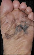
|

|
Other histopathologic types are:
- Mucosal melanoma; When melanoma occurs on mucous membranes.
- Desmoplastic melanoma
- Melanoma with small nevus-like cells
- Melanoma with features of a Spitz nevus
- Uveal melanoma
- Vaginal melanoma
- Polypoid melanoma, a subclass of nodular melanoma.
In situ or invasive
A melanoma in situ has not invaded beyond the basement membrane, whereas an invasive melanoma has spread beyond it.
Some histopathological types of melanoma are inherently invasive, including nodular melanoma and lentigo maligna melanoma, where the in situ counterpart to lentigo maligna melanoma is lentigo maligna. Lentigo maligna is sometimes classified as a very early melanoma, and sometimes a precursor to melanoma.
Superficial spreading melanomas and acral lentiginous melanomas can be either in situ or invasive, but acral lentiginous melanomas are almost always invasive.
Staging
Further context on cancer staging is available at TNM.

Metastatic melanomas can be detected by X-rays, CT scans, MRIs, PET and PET/CTs, ultrasound, LDH testing and photoacoustic detection. However, there is lack of evidence in the accuracy of staging of people with melanoma with various imaging methods.
Melanoma stages according to AJCC, 8th edition:
- TX: Primary tumor thickness cannot be assessed (such as diagnosis by curettage)
- T0: No evidence of primary tumor (such as unknown primary or completely regressed melanoma)
| Stage | T category | Thickness | Ulceration |
|---|---|---|---|
| Stage 0 | Melanoma in situ | ||
| Stage I | T1a | Less than 0.8 mm | No |
| T1b | Less than 0.8 mm | No | |
| >0.8 to 1.0 mm | Yes | ||
| T2a | >1.0 to 2.0 mm | No | |
| Stage II | T2b | >1.0 to 2.0 mm | Yes |
| T3a | >2.0 to 4.0 mm | No | |
| T3b | >2.0 to 4.0 mm | Yes | |
| T4a | >4.0 mm | No | |
| T4b | >4.0 mm | Yes | |
Stage 1 and 2 require an N (lymph node) class of:
- N0 – No regional metastases.
| Stage | N category | Number of tumor-involved regional lymph nodes | Presence of in-transit, satellite, and/or microsatellite metastases |
|---|---|---|---|
| N/A | NX | Regional nodes not assessed (such as sentinel lymph node biopsy not performed, or regional nodes previously removed for another reason) | |
| Stage III | N1 | One involved lymph node, or any number of in-transit, satellite, and/or microsatellite metastases with no tumor-involved nodes. | |
| N1a | One clinically occult (that is, detected by sentinel node biopsy) | No | |
| N1b | One clinically detected | No | |
| N1c | No regional lymph node disease | Yes | |
| N2 | Two or 3 tumor‐involved nodes or any number of in‐transit, satellite, and/or microsatellite metastases with one tumor‐involved node | ||
| N2a | Two or 3 clinically occult (that is, detected by sentinel node biopsy) | No | |
| N2b | Two or 3, at least one of which was clinically detected | No | |
| N2c | One clinically occult or clinically detected | Yes | |
| N3 | Four or more tumor‐involved nodes or any number of in‐transit, satellite, and/or microsatellite metastases with 2 or more tumor‐involved nodes, or any number of matted nodes without or with in‐transit, satellite, and/or microsatellite metastases | ||
| N3a | Four or more clinically occult (that is, detected by sentinel node biopsy) | No | |
| N3b | Four or more, at least one of which was clinically detected, or the presence of any number of matted nodes | No | |
| N3c | Two or more clinically occult or clinically detected and/or presence of any number of matted nodes | Yes | |
Stage 1, 2 and 3 require an M (metastasis status) of:
- M0: No evidence of distant metastasis
| Stage | M category | Anatomic site | lactate dehydrogenase (LDH) level |
|---|---|---|---|
| Stage IV | M1 | Evidence of distant metastasis | |
| M1a | Distant metastasis to skin, soft tissue including muscle, and/or non-regional lymph node | Not recorded or unspecified | |
| M1a(0) | Not elevated | ||
| M1a(1) | Elevated | ||
| M1b | Distant metastasis to lung with or without metastasis at M1a sites | Not recorded or unspecified | |
| M1b(0) | Not elevated | ||
| M1b(1) | Elevated | ||
| M1c | Distant metastasis to non‐CNS visceral sites, with or without metastasis to M1a or M1b sites | Not recorded or unspecified | |
| M1c(0) | Not elevated | ||
| M1c(1) | Elevated | ||
| M1d | Distant metastasis to CNS, with or without metastasis to M1a, M1b, or M1c sites | Not recorded or unspecified | |
| M1d(0) | Not elevated | ||
| M1d(1) | Elevated | ||
Older systems include "Clark level" and "Breslow's depth", quantifying microscopic depth of tumor invasion.

Laboratory
Lactate dehydrogenase (LDH) tests are often used to screen for metastases, although many patients with metastases (even end-stage) have a normal LDH; extraordinarily high LDH often indicates metastatic spread of the disease to the liver.
It is common for patients diagnosed with melanoma to have chest X-rays and an LDH test, and in some cases CT, MRI, PET, and/or PET/CT scans. Although controversial, sentinel lymph node biopsies and examination of the lymph nodes are also performed in patients to assess spread to the lymph nodes. A diagnosis of melanoma is supported by the presence of the S-100 protein marker.
HMB-45 is a monoclonal antibody that reacts against an antigen present in melanocytic tumors such as melanomas. It is used in anatomic pathology as a marker for such tumors. The antibody was generated to an extract of melanoma. It reacts positively against melanocytic tumors but not other tumors, thus demonstrating specificity and sensitivity. The antibody also reacts positively against junctional nevus cells but not intradermal nevi, and against fetal melanocytes but not normal adult melanocytes.
HMB-45 is nonreactive with almost all non-melanoma human malignancies, with the exception of rare tumors showing evidence of melanogenesis (e.g., pigmented schwannoma, clear cell sarcoma) or tumors associated with tuberous sclerosis complex (angiomyolipoma and lymphangiomyoma).
Prevention
There is no evidence to support or refute adult population screening for malignant melanoma.
Ultraviolet radiation
Minimizing exposure to sources of ultraviolet radiation (the sun and sunbeds), following sun protection measures and wearing sun protective clothing (long-sleeved shirts, long trousers, and broad-brimmed hats) can offer protection.
Using artificial light for tanning was once believed to help prevent skin cancers, but it can actually lead to an increased incidence of melanomas.
UV nail lamps, which are used in nail salons to dry nail polish, are another common and widespread source of UV radiation that could be avoided. Although the risk of developing skin cancer through UV nail lamp use is low, it is still recommended to wear fingerless gloves and/or apply SPF 30 or greater sunscreen to the hands before using a UV nail lamp.
The body uses UV light to generate vitamin D so there is a need to balance getting enough sunlight to maintain healthy vitamin D levels and reducing the risk of melanoma; it takes around a half hour of sunlight for the body to generate its vitamin D for the day and this is about the same amount of time it takes for fair-skinned people to get a sunburn. Exposure to sunlight can be intermittent instead of all at one time.
Sunscreen
Sunscreen appears to be effective in preventing melanoma. In the past, use of sunscreens with a sun protection factor (SPF) rating of 50 or higher on exposed areas were recommended; as older sunscreens more effectively blocked UVA with higher SPF. Currently, newer sunscreen ingredients (avobenzone, zinc oxide, and titanium dioxide) effectively block both UVA and UVB even at lower SPFs. Sunscreen also protects against squamous cell carcinoma, another skin cancer.
Concerns have been raised that sunscreen might create a false sense of security against sun damage.
Medications
A 2005 review found tentative evidence that statin and fibrate medication may decrease the risk of melanoma. A 2006 review however did not support any benefit.
Treatment

Confirmation of the clinical diagnosis is done with a skin biopsy. This is usually followed up with a wider excision of the scar or tumor. Depending on the stage, a sentinel lymph node biopsy may be performed. Controversy exists around trial evidence for sentinel lymph node biopsy; with unclear evidence of benefit as of 2015. Treatment of advanced malignant melanoma is performed from a multidisciplinary approach.
Surgery
Excisional biopsies may remove the tumor, but further surgery is often necessary to reduce the risk of recurrence. Complete surgical excision with adequate surgical margins and assessment for the presence of detectable metastatic disease along with short- and long-term followup is standard. Often this is done by a wide local excision (WLE) with 1–2 cm (0.4–0.8 in) margins. Melanoma-in-situ and lentigo malignas are treated with narrower surgical margins, usually 0.2–0.5 cm (0.1–0.2 in). Many surgeons consider 0.5 cm (0.2 in) the standard of care for standard excision of melanoma-in-situ, but 0.2 cm (0.1 in) margin might be acceptable for margin controlled surgery (Mohs surgery, or the double-bladed technique with margin control). The wide excision aims to reduce the rate of tumor recurrence at the site of the original lesion. This is a common pattern of treatment failure in melanoma. Considerable research has aimed to elucidate appropriate margins for excision with a general trend toward less aggressive treatment during the last decades. A 2009 meta-analysis of randomized controlled trials found a small difference in survival rates favoring wide excision of primary cutaneous melanomas, but these results were not statistically significant.
Mohs surgery has been reported with cure rate as low as 77% and as high as 98.0% for melanoma-in-situ. CCPDMA and the "double scalpel" peripheral margin controlled surgery is equivalent to Mohs surgery in effectiveness on this "intra-epithelial" type of melanoma.
Melanomas that spread usually do so to the lymph nodes in the area of the tumor before spreading elsewhere. Attempts to improve survival by removing lymph nodes surgically (lymphadenectomy) were associated with many complications, but no overall survival benefit. Recently, the technique of sentinel lymph node biopsy has been developed to reduce the complications of lymph node surgery while allowing assessment of the involvement of nodes with tumor.
Biopsy of sentinel lymph nodes is a widely used procedure when treating cutaneous melanoma.
Neither sentinel lymph node biopsy nor other diagnostic tests should be performed to evaluate early, thin melanoma, including melanoma in situ, T1a melanoma or T1b melanoma ≤ 0.5mm. People with these conditions are unlikely to have the cancer spread to their lymph nodes or anywhere else and have a 5-year survival rate of 97%. Because of these considerations, sentinel lymph node biopsy is considered unnecessary health care for them. Furthermore, baseline blood tests and radiographic studies should not be performed only based on identifying this kind of melanoma, as there are more accurate tests for detecting cancer and these tests have high false-positive rates. To potentially correct false positives, gene expression profiling may be used as auxiliary testing for ambiguous and small lesions.
Sentinel lymph node biopsy is often performed, especially for T1b/T2+ tumors, mucosal tumors, ocular melanoma and tumors of the limbs. A process called lymphoscintigraphy is performed in which a radioactive tracer is injected at the tumor site to localize the sentinel node(s). Further precision is provided using a blue tracer dye, and surgery is performed to biopsy the node(s). Routine hematoxylin and eosin (H&E) and immunoperoxidase staining will be adequate to rule out node involvement. Polymerase chain reaction (PCR) tests on nodes, usually performed to test for entry into clinical trials, now demonstrate that many patients with a negative sentinel lymph node actually had a small number of positive cells in their nodes. Alternatively, a fine-needle aspiration biopsy may be performed and is often used to test masses.
If a lymph node is positive, depending on the extent of lymph node spread, a radical lymph node dissection will often be performed. If the disease is completely resected, the patient will be considered for adjuvant therapy. Excisional skin biopsy is the management of choice. Here, the suspect lesion is totally removed with an adequate (but minimal, usually 1 or 2 mm) ellipse of surrounding skin and tissue. To avoid disruption of the local lymphatic drainage, the preferred surgical margin for the initial biopsy should be narrow (1 mm). The biopsy should include the epidermal, dermal, and subcutaneous layers of the skin. This enables the histopathologist to determine the thickness of the melanoma by microscopic examination. This is described