Thymoma

Thymoma is a thymic epithelial neoplasm (TEN; see this term), a rare malignancy that arises from the epithelium of the thymic gland.
Epidemiology
It is the most common form of TEN and has an annual incidence of approximately 1/769,000. The male to female ratio is 1:1.4. Thymomas usually occur between the age of 30 and 70 years (with a median age of 50 years) but in some rare cases they can also occur during childhood.
Clinical description
While half of the patients are asymptomatic, the other half present with chest related symptoms such as dyspnea, chest pain, upper respiratory infection, fatigue, weight loss, and cough or pneumonia. Thymomas are often associated with myasthenia gravis (see this term), an autoimmune disorder that manifests with diplopia, ptosis, dysphagia, and weakness. Some patients may suffer from other autoimmune diseases such as systemic lupus erythematosus (SLE; see this term) and rheumatoid arthritis, hematologic syndromes such as red cell anaplasia and erythrocytosis, and other chronic diseases such as hypertension, diabetes mellitus, renal insufficiency and coronary artery disease. A history of second tumor may be present in some patients.
Etiology
Etiology is unknown.
Diagnostic methods
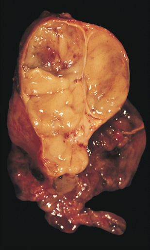
Diagnosis is based on clinical findings, radiological studies and pathologic examination of resected tissues. Chest X-rays may reveal a widened mediastinum or loss of the normal anterior clear space. Computed tomography, magnetic resonance imaging (MRI), magnetic resonance angiography (angio-MRI) and/or positron emission tomography (PET) can be indicated to further evaluate the lesion. Tissue biopsy, usually performed by CT- or ultrasound-guided percutaneous needle-biopsy, is necessary for diagnosis. Thymomas are currently divided according to the World Health Organization (WHO) into four histological subclasses: types A, B, AB, and C. Type A tumors are composed of spindle or oval epithelial cells. Type B tumors consist of round or polygonal epithelial cells and are divided into three subtypes (B1, B2, and B3) based on their various proportions of background lymphocytes and increase in cytologic atypia of the epithelial cells. Type AB tumors consist of type A thymoma features with a variably dense lymphocytic component. Type C tumors are characterized by cytologic atypia and loss of the organotypical characteristics of the thymus; the latter term is synonymous with thymic carcinoma (see this term). On cut section, the tumors are nodular and gray-white, can be multi-cystic and can contain calcifications or hemorrhage. Most of them are encapsulated and some may be invasive.
Differential diagnosis
Differential diagnoses include lymphoma, germ-cell tumors (see these terms), and metastatic cancers.
Management and treatment
In early stages, treatment consists of complete surgical excision (usually performed by median sternotomy). In advanced stages (stage II according to Masaoka staging system) and in high-risk histologic subtypes (B3), surgery is accompanied by additional adjuvant therapy (consisting of post-operative radiotherapy) or neoadjuvant therapy. Total thymectomy usually relieves myasthenia gravis symptoms.