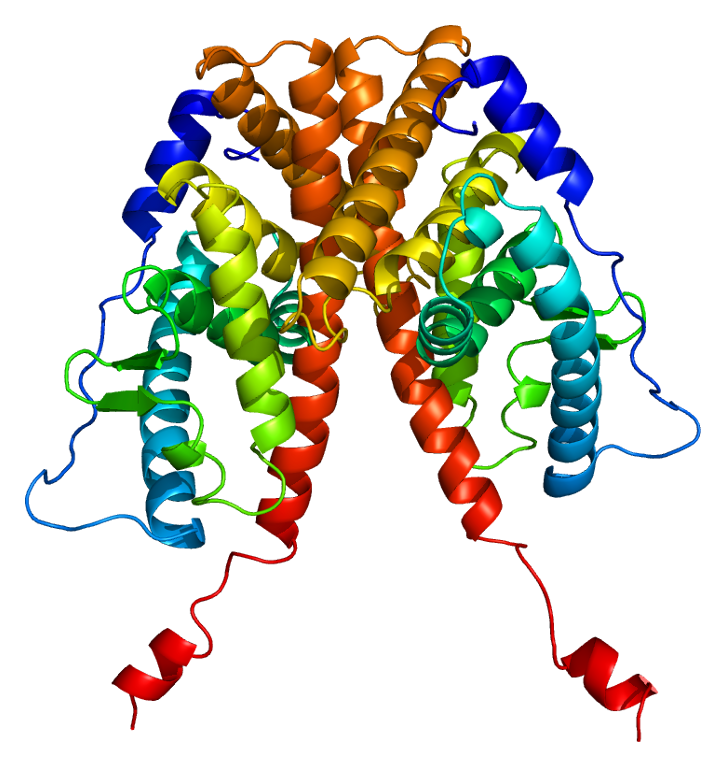
Estrogen Resistance

A number sign (#) is used with this entry because of evidence that estrogen resistance (ESTRR) is caused by homozygous mutation in the alpha estrogen receptor gene (ESR1; 133430) on chromosome 6q25.
DescriptionEstrogen resistance (ESTRR) is characterized by absence of puberty with elevated estradiol and gonadotropic hormones, as well as markedly delayed bone maturation. Female patients show absent breast development, small uterus, and enlarged multicystic ovaries; male patients may show small testes (Bernard et al., 2017). Some patients exhibit continued growth into adulthood (Smith et al., 1994).
Clinical FeaturesSmith et al. (1994) described a 28-year-old man with estrogen resistance. He was 204 cm tall and had incomplete epiphyseal closure, with a history of continued linear growth into adulthood despite otherwise normal pubertal development. He was normally masculinized and had bilateral axillary acanthosis nigricans. Serum estradiol and estrone concentrations were elevated, and serum testosterone concentrations were normal. Serum follicle-stimulating hormone and luteinizing hormone concentrations were increased. Glucose tolerance was impaired and hyperinsulinemia was present. Bone mineral density of the lumbar spine was 3.1 SD below the mean for age-matched normal women; there was no biochemical evidence of increased bone turnover. Administration of estrogen had no detectable effect. Smith et al. (1994) concluded that estrogen is important for bone maturation and mineralization in men as well as in women.
Sudhir et al. (1997) presented further clinical and laboratory findings from the man originally reported by Smith et al. (1994). Electron-beam CT scanning of the patient at age 31 years showed evidence of early atherosclerosis in the left anterior descending coronary artery. Abnormal serum lipid concentrations were thought to reflect estrogen insensitivity. The authors concluded that the absence of functional estrogen receptors might be a novel risk factor for coronary artery disease in men.
Smith et al. (2008) performed a follow-up of the propositus with a homozygous nonsense mutation in ESR1 (see MOLECULAR GENETICS) originally reported by Smith et al. (1994), combined with an assessment of the effect of heterozygosity on spine density and adult height in members of his extended kindred. Patient bone biopsy revealed marked osteopenia, low trabecular volume, decreased thickness, normal trabecular number, and low activation frequency. Trabecular and cortical volumetric bone densities were markedly lower than controls. Heterozygous mutation carriers had mean spine aBMD Z scores that were significantly less than zero (p = 0.003), but spine aBMD Z scores were similar among carrier and noncarrier family members. The authors concluded that homozygous ESR1 disruption markedly affects bone growth, mineral content, and structure, but not periosteal circumference, whereas heterozygosity for ESR1 mutations appears not to impair areal spine BMD.
Quaynor et al. (2013) studied an 18-year-old woman who presented at 15 years of age with absent breast development, primary amenorrhea, and intermittent lower abdominal pain. Examination revealed Tanner stage 1 breast development and Tanner stage 4 pubic hair, and she also had severe facial acne. She was found to have markedly elevated serum levels of estrogens, with a plasma estradiol level 10 times that of the normal value, and had a bone age of 11 to 12 years at a chronologic age of 15.5 years. Ultrasonography revealed a small uterus with no clearly identifiable endometrial stripe and markedly enlarged multicystic ovaries. Growth velocity indicated the lack of an estrogen-induced growth spurt at the time of puberty. At the age of 17.75 years, plain x-rays of her left hand showed a bone age of 13.5 years and open radial and ulnar epiphyses. MRI of the brain and pituitary was normal. She had no breast development after 5 months of receiving oral estrogen, and her normal serum levels of sex-hormone-binding globulin, corticosteroid-binding globulin, thyroxine-binding globulin, prolactin and triglycerides, all of which are known to be increased by estrogen, provided further evidence of estrogen resistance. Unlike the male patient with estrogen resistance reported by Smith et al. (1994), who had hyperinsulinemia, she had normal levels of fasting glucose, insulin, and glycated hemoglobin, and a normal HOMA-IR, indicating no impaired glucose tolerance.
Bernard et al. (2017) reported 3 sibs from a consanguineous Algerian family with estrogen resistance. The proband was a 25-year-old 46,XX woman who presented with primary amenorrhea and absent breast development; pelvic ultrasonography showed bilateral enlarged multicystic ovaries and small uterus with thin endometrium. She had an extremely high 17-beta-estradiol level as well as elevated gonadotropins. Her 21-year-old 46,XX sister also showed no breast development and had primary amenorrhea. In addition, their 18-year-old 46,XY brother had Tanner stage I gonadal development, with a cryptorchid right testis and left testis volume of less than 1 cm. All 3 patients exhibited markedly delayed bone maturation. Their first-cousin parents had normal pubertal development at 15 years of age, and an unaffected sister had normal puberty with menarche at age 14 years.
Molecular GeneticsIn a 28-year-old man with estrogen resistance, Smith et al. (1994) performed single-strand conformation polymorphism analysis of the ESR1 gene (133430) and observed a variant banding pattern in exon 2. Direct sequencing revealed a homozygous nonsense mutation (R157X; 133430.0002). Both parents were heterozygous carriers of the R157X mutation, and pedigree analysis showed that they were related as second cousins; 3 sisters were also heterozygous.
In an 18-year-old woman with estrogen resistance, Quaynor et al. (2013) identified homozygosity for a missense mutation in the ESR1 gene (Q375H; 133430.0006). The patient had been adopted, and parental DNA samples were not available.
Clegg and Palmer (2013) noted that the Esr1 -/AA knockin mouse model recapitulates the phenotype of the female patient studied by Quaynor et al. (2013), in that they have hypoplastic mammary glands, anovulation, and altered steroidogenesis, but have normal body weight, adiposity, locomotor activity, and glucose homeostasis. These findings supported the proposal by Quaynor et al. (2013) that nonclassical regulation of ESR1 is sufficient to protect against the obesity-metabolic syndrome phenotype associated with total loss of activity of ESR1, but is not sufficient to rescue the infertility phenotype.