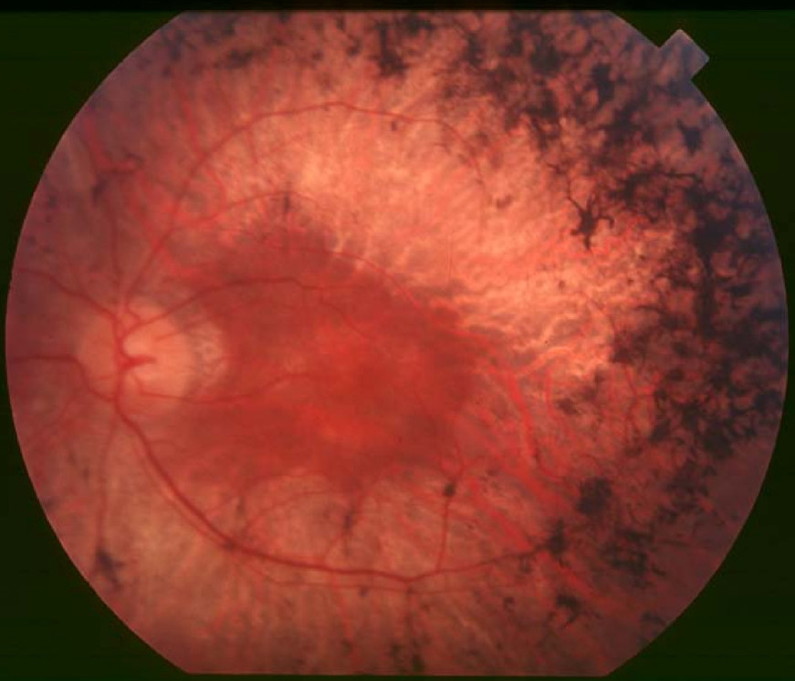
Retinitis Pigmentosa 9

A number sign (#) is used with this entry because of evidence that retinitis pigmentosa-9 (RP9) is caused by heterozygous mutation in the RP9 gene (607331) on chromosome 7p14. One such patient has been reported.
For a phenotypic description and a discussion of genetic heterogeneity of retinitis pigmentosa, see 268000.
DescriptionAutosomal dominant retinitis pigmentosa (ADRP) is characterized by a typical fundus appearance, narrowed retinal vessels, and changes in the electrophysiological responses of the eye. Early signs are night blindness and constriction of the visual fields with a variable ages of onset (summary by Jay et al., 1992).
Clinical FeaturesJay et al. (1992) described a 9-generation family (family N) with autosomal dominant retinitis pigmentosa. Using census records, hospital records from the Moorfields Eye Hospital, and telephone interviews with relatives, the authors determined the age of onset and visual outcome of 73 family members. Moore et al. (1993) performed detailed eye examinations of 10 members of the family described by Jay et al. (1992) (family 2 in Moore et al., 1993). Affected individuals had a variable age of onset of symptoms: under 10 years in 2 patients, between 11 and 20 years in 3 patients, and over 21 years in 5 patients. Six of the 10 patients had either macular atrophy or edema and half had cataracts. Psychophysical testing and electroretinography showed variation in the severity of the disease that was not determined by age.
InheritanceThe transmission pattern of retinitis pigmentosa in the family described by Jay et al. (1992) and Moore et al. (1993) was consistent with autosomal dominant inheritance with incomplete penetrance.
MappingIn 2 large families with autosomal dominant retinitis pigmentosa in which linkage to rhodopsin (180380) had been excluded, Bashir et al. (1992) reported exclusion data also for chromosomes 6 and 8. Bashir et al. (1992) concluded that there is a form of autosomal dominant retinitis pigmentosa in addition to the 3 varieties that had been demonstrated by linkage or other studies: the rhodopsin-related form on 3q (RP4; 613731), the peripherin (PRPH2; 179605)-related form on 6p (RP7; 608133), and the form linked to 8p (RP1, 180100, caused by mutations in ORP1 603937).
In the British family originally reported by Jay et al. (1992), Inglehearn et al. (1993) demonstrated linkage of autosomal dominant retinitis pigmentosa to 2 microsatellite markers on 7p: D7S435, which had previously been localized to 7p15.1-p13, and D7S460, which mapped to within 2 cM of D7S435 with a lod score of 12.15. Multipoint analysis gave a maximum lod score of 8.22 for retinitis pigmentosa in this family with the 2 markers. They referred to the entity as adRP7.
Jordan et al. (1993) studied a Spanish family in which positive 2-point lod scores were obtained with 15 markers. Multipoint analyses using a subset of these markers gave a lod score of 7.51, maximizing at D7S480. These data provided evidence for an adRP gene on 7q; see 180105. They excluded linkage to the markers closely linked to RP in the British pedigree of Inglehearn et al. (1993). The severity of adRP in the Spanish family was greater than that in the British family, members of which showed a much later onset of symptoms. The Spanish family became aware of symptoms at a mean age of 12.9 years.
Inglehearn et al. (1994) showed by linkage analysis that RP9 is separate from dominant cystoid macular dystrophy (153880), which also maps to 7p but at a distance of approximately 10 cM from RP9. Keen et al. (1995) reported a 4.8-Mb YAC contig spanning the RP9 locus.
Molecular GeneticsKeen et al. (2002) described a previously uncharacterized human gene (RP9; 607331) mapping to the RP9 critical interval at 7p14.2. A missense mutation (H137L; 607331.0001) was identified in affected members and obligate carriers in the British family with linkage to 7p originally described by Inglehearn et al. (1993). Another missense mutation (D170G; 607331.0002) in the RP9 gene was identified in a single retinitis pigmentosa patient by screening a panel of 300 dominant, recessive, and genetically undefined retinitis pigmentosa patients. The phenotype of this patient was consistent with that described for the RP9 family. The function of the RP9 gene was unknown and the pathogenic mechanism remained to be determined.
Maita et al. (2004) used in vitro and in vivo splicing assays to examine the H137L (607331.0001) and D170G (607331.0002) mutant forms of PAP1. The H137L mutant had no effect on splicing activity compared with that of wildtype PAP1, calling into question the pathogenicity of this mutation. The D170G mutant showed a defect in splicing activity and a decreased proportion of phosphorylated PAP1.
Sullivan et al. (2006) used Sanger sequencing to screen 200 families with autosomal dominant retinitis pigmentosa and did not find any pathogenic mutations in PAP1.
Exclusion Studies
Because expanded tracts of (CAG)n had been found in certain genes as the cause of neurodegenerative disease, Keen et al. (1997) sought evidence of (CAG)n expansions as the cause of disease in a panel of 8 autosomal dominant retinitis pigmentosa pedigrees, including families known to map to the RP9, RP11 (600138), and RP13 (600059) loci, using the technique known as repeat expansion detection (RED). In the family studies no evidence of such expanded repeats was uncovered.