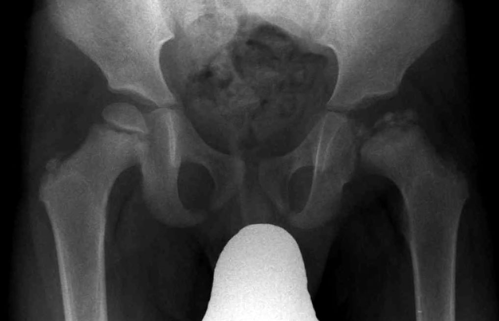
Legg-Calvé-Perthes Disease

A rare disorder characterized by uni- or bilateral avascular necrosis (AVN) of the femoral head in children.
Epidemiology
Reported annual incidences vary greatly, from 1/250,000 in Hong Kong and 1/18,000 in the UK, to 1/3,500 in the Faroe Islands. Legg-Calvé-Perthes disease (LCPD) affects children between 2 and 12 years of age, but it is more prevalent among children of 5-6 years, and more common in boys.
Clinical description
The initial symptoms are usually a limping gait, pain in the hip, thigh or knee, and a reduced range of hip motion. Later in the disease course, leg length discrepancy, as well as atrophy of musculature around the hip can be observed. The active phase of the disease can last for several years, and during this phase the femoral head becomes partially or completely necrotic and gradually deformed. This is followed by new bone formation (re-ossification) in the epiphysis and eventual healing. The final deformity can vary from a nearly normal joint configuration to an extensive deformation with severe flattening and subluxation of the femoral head, broadening of the femoral neck, and a deformed and dysplastic acetabulum, which in turn can lead to early-onset osteoarthritis.
Etiology
The etiology of LCPD remains obscure. It is generally accepted that one or more infarctions of the femoral head due to interruption of vascular supply eventually cause the deformity, however, there are several theories concerning the cause of this interruption. Several contributory factors have also been suggested: delayed skeletal maturity, impaired and disproportionate growth, short stature, low birth weight, social and economic deprivation and trauma, as well as an association with congenital anomalies. It has also been proposed that coagulation system disorders could cause thrombophilia and/or hypofibrinolysis and lead to thrombotic venous occlusion with subsequent AVN of the femoral head in children. Mutations in the COL2A1 gene (12q12-q13.2) have recently been identified in familial cases of AVN of the femoral head (see this term) and LCPD.
Diagnostic methods
Diagnosis is made by conventional radiography in frontal and lateral projections. Scintigraphy and ultrasound can be of value in selected cases and MRI can be useful in the early stages of the disease to distinguish LCPD from other hip disorders.
Differential diagnosis
Differential diagnoses include Meyers dysplasia, multiple epiphyseal dysplasia and spondyloepiphyseal dysplasia (see these terms).
Management and treatment
The main aim of treatment is to contain the femoral head within the acetabulum, either using an abduction brace or through surgical interventions (femoral or pelvic osteotomy). A recent study suggested that femoral osteotomy gives significantly better results than treatment with braces (specifically the Scottish Rite abduction orthosis).
Prognosis
Prognosis is variable and several factors are of prognostic importance, such as the extent of femoral head necrosis and residual deformity. The more deformed the femoral head is during healing, the greater the risk of osteoarthritis later in life. Total hip replacement in early adulthood may be required in some cases. A younger age at diagnosis is generally accepted to be associated with a better outcome.