Retinitis Pigmentosa 66

A number sign (#) is used with this entry because of evidence that retinitis pigmentosa-66 (RP66) is caused by homozygous mutation in the RBP3 gene (180290) on chromosome 10q11. One such family has been reported.
For a general phenotypic description and a discussion of genetic heterogeneity of retinitis pigmentosa (RP), see 268000.
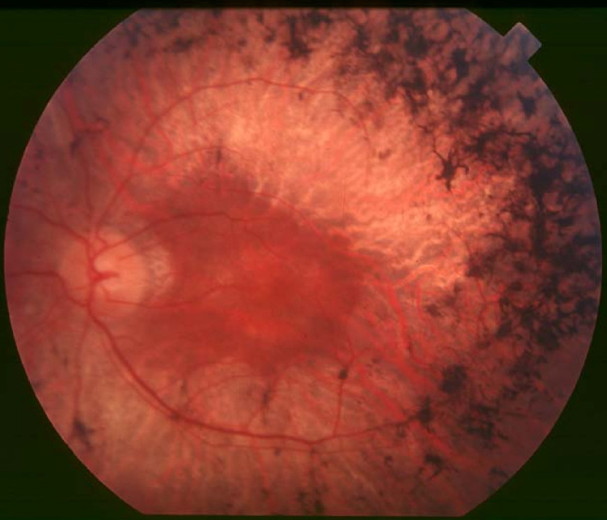
Clinical FeaturesDen Hollander et al. (2009) studied a consanguineous Italian family in which 3 brothers and a sister had retinitis pigmentosa; 2 of the brothers underwent detailed clinical evaluation, which demonstrated a wide range of severity in this family. The 42-year-old brother reported loss of central vision and onset of night blindness at 32 years of age, whereas his older brother had loss of central vision at age 60 with no night deficiency even at 67 years of age. The older brother had visual acuities of 20/60 and 20/80 and normal color vision, whereas the younger had acuities of 20/200 bilaterally, with a tritan axis of confusion on the Farnsworth-D-15 panel. The authors noted that the older brother had increased macular thickness due to cystoid macular edema and the younger brother had reduced central retinal thickness on optical coherence tomography as possible explanations for their decreased acuities. Visual fields showed marked constriction with central scotoma in both patients, although the visual fields were more severely diminished in the younger brother. Both patients showed clumped bone spicule pigment around the periphery and attenuated retinal vessels; the younger patient had waxy pallor of the optic disc, whereas the older brother had normal color, with a large area of atrophy temporal to each disc. Both brothers had posterior subcapsular cataracts. Electroretinograms (ERGs) in both patients showed profound loss of rod and cone function, with ERG amplitudes so reduced that they could be detected only by computer averaging and narrow band-pass filtering. The cone amplitudes in the younger brother were smaller at age 46 than those in his older brother at age 67; both had delayed cone implicit times that were consistent with progressive disease. Multiple follow-up examinations in the younger brother revealed much slower rates of field and ERG loss than average reported rates for untreated RP. The other 2 sibs were reported to have funduscopy findings compatible with RP and very reduced ERG responses that were virtually nondetectable without computer averaging. Both parents, who died later in life, and 1 brother were unaffected.
MappingIn a consanguineous Italian family in which 4 sibs had retinitis pigmentosa, den Hollander et al. (2009) performed homozygosity mapping with SNP microarrays that revealed only 1 homozygous shared region, a 40-Mb region on chromosome 10 spanning 3,780 SNPs that segregated completely with disease in the family. Recombination events narrowed the critical interval to 9 Mb between SNPs rs2460551 and rs7898315.
Molecular GeneticsIn a consanguineous Italian family with retinitis pigmentosa mapping to chromosome 10, den Hollander et al. (2009) analyzed 3 candidate genes and identified a missense mutation in the RBP3 gene (D1080N; 180290.0001) that segregated with disease and was not found in 116 Italian controls or 94 controls of mixed North American ancestry. Analysis of RBP3 in 395 additional unrelated patients with recessive or sporadic RP and in 680 patients with other forms of hereditary retinal degeneration revealed no mutations.
Using D-HPLC and direct sequencing, Ksantini et al. (2010) screened the RBP3 gene in 134 patients with autosomal recessive or sporadic RP and 82 patients with other retinal dystrophies, but did not find any pathogenic mutations. The authors concluded that mutations in RPB3 occur rarely in inherited retinal dystrophies.