Retinitis Pigmentosa 10

A number sign (#) is used with this entry because of evidence that retinitis pigmentosa-10 (RP10) is caused by heterozygous mutation in the IMPDH1 gene (146690) on chromosome 7q32.
Heterozygous mutation in the IMPDH1 gene can also cause Leber congenital amaurosis-11 (LCA11; 613837).
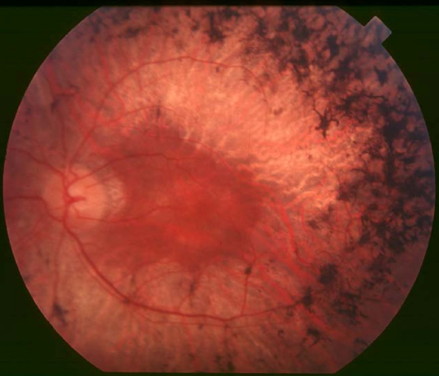
DescriptionRetinitis pigmentosa-10 (RP10) is characterized in most patients by early onset and rapid progression of ocular symptoms, beginning with night blindness in childhood, followed by visual field constriction. Some patients experience an eventual reduction in visual acuity. Funduscopy shows typical changes of RP, including optic disc pallor, retinal vascular attenuation, and bone-spicule pattern of pigmentary deposits in the retinal midperiphery. Electroretinography demonstrates equal reduction in rod and cone responses (Jordan et al., 1993; Bowne et al., 2002; Bowne et al., 2006).
For a general phenotypic description and a discussion of genetic heterogeneity of retinitis pigmentosa, see 268000.
Clinical FeaturesJordan et al. (1993) reported a Spanish family with retinitis pigmentosa showing relatively early onset of symptoms (mean age of onset, 12.9 years).
Coussa et al. (2015) reported a 40-year-old French Canadian man with retinitis pigmentosa. The patient stated that he had decreased peripheral vision loss, color vision fluctuation, progressively worsening nyctalopia, and central vision decrease since early childhood. At examination, his visual acuity was 20/400 OD and 20/200 OS. The posterior pole was remarkable for classic bone spicules, severe fundus and optic nerve pallor, atrophic disc, attenuation and straightening of the blood vessels, and bull's-eye maculopathy with extensive concentric areas of clumped hyperpigmentation. FAF imaging showed central hypopigmented confluent islands surrounded by a hyperpigmented crescent. OCT was remarkable for severe foveal dipping, outer retinal tissue thinning, and cystic changes. The choroidal layer had diffuse cystic lesions also.
MappingIn a Spanish family with early-onset retinitis pigmentosa, Jordan et al. (1993) found linkage to markers on 7q and excluded linkage to markers on 7p where a gene for adRP had been located by linkage by Inglehearn et al. (1993); see 180104.
In a large American family with late-onset autosomal dominant RP, McGuire et al. (1995) used microsatellite markers to demonstrate linkage to 7q. A maximum 2-point lod score of 5.3 at 0% recombination was found with D7S514. The linkage studies provided strong evidence that RP10 is located in the 7q31-q35 region.
In a second Spanish family with adRP, Millan et al. (1995) demonstrated linkage to 7q31-q35 with a maximum lod score of 3.01 for D7S480 by multipoint analysis.
McGuire et al. (1996) combined linkage results from the original Spanish family and an unrelated American family to assign the disease locus to a 5-cM interval on 7q. Based on extensive physical mapping of this region, the genetic interval was found to be contained fully within a segment of approximately 5 Mb on a well-defined YAC contig.
Molecular GeneticsBy linkage mapping, Bowne et al. (2002) identified 2 American families with the RP10 form of adRP and used these families to reduce the linkage interval to 3.45 Mb between the flanking markers D7S686 and RP-STR8. Ten retinal transcripts were identified among 54 independent genes within the candidate region, including IMPDH1. DNA sequencing of affected individuals from 3 RP10 families, 2 from the US and 1 from the UK, revealed an asp226-to-asn substitution in IMPDH1 (146690.0001). Asp226 is highly evolutionarily conserved among IMPDH genes, suggesting that this mutation may be highly deleterious. Another IMPDH1 substitution, val268 to ile (146690.0002), was observed in one of a cohort of 60 adRP families but not in controls. IMPDH1 is a ubiquitously expressed enzyme, functioning as a homotetramer, which catalyzes the rate-limiting step in de novo synthesis of guanine nucleotides. As such, it may play an important role in cyclic nucleotide metabolism within photoreceptors.
Kennan et al. (2002) used microarray analysis to compare retinal transcript levels between wildtype mice and those with a targeted disruption of the rhodopsin gene (180380), designated Rho -/-. The IMPDH1 gene was identified among a series of transcripts present at reduced levels. Mutational screening of DNA from the Spanish adRP family reported by Jordan et al. (1993) revealed an arg224-to-pro substitution (R224P; 146690.0003) cosegregating with the disease phenotype. Arg224 is conserved among species, and the substitution was not present in a European control population.
Among 60 French Canadian patients with RP, Coussa et al. (2015) identified a causal mutation in 24 patients, one of whom had a heterozygous mutation in the IMPDH1 gene (Q318H; 146690.0006).