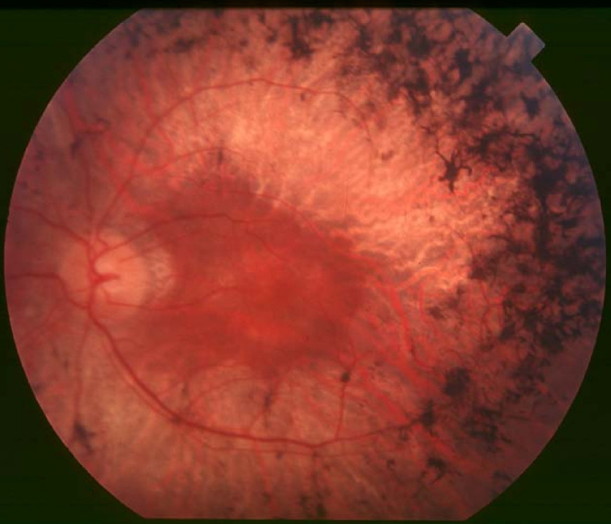
Retinitis Pigmentosa 12

A number sign (#) is used with this entry because of evidence that retinitis pigmentosa-12 (RP12) is caused by homozygous or compound heterozygous mutations in the CRB1 gene (604210) on chromosome 1q31.
Homozygous or compound heterozygous mutation in CRB1 can also cause a more severe retinal dystrophy, Leber congenital amaurosis (LCA8; see 604210).
For a phenotypic description and a discussion of genetic heterogeneity of retinitis pigmentosa, see 268000.
Clinical FeaturesHeckenlively (1982) described 5 patients with retinitis pigmentosa of probable autosomal recessive inheritance who showed relative preservation of retinal pigment epithelium adjacent to and under retinal arterioles and hypermetropia (RP patients tend to be myopic). Affected sibs and parental consanguinity were noted.
Bleeker-Wagemakers et al. (1992) reported a consanguineous Dutch family of 90 members from a genetically isolated population in the north of the Netherlands. Affected members (approximately 40) presented with an early-onset form of retinitis pigmentosa, starting with night blindness before the age of 3 years. Nonrecordable electroretinograms (ERGs) and concentric visual field loss in the first decade were followed in most cases by macular involvement and decreased visual acuity before age 15. Visual acuity at age 30 was 0.2 or less. At funduscopy, pigmentary depositions and atrophy of retinal pigment epithelium (RPE) were seen. See also Humphries et al. (1992).
Van den Born et al. (1994) observed clinical heterogeneity within the pedigree reported by Bleeker-Wagemakers et al. (1992). Only some affected members displayed retinitis pigmentosa with characteristic preserved paraarteriolar RPE (PPRPE).
Benayoun et al. (2009) studied a consanguineous family in which 4 of 9 offspring had severe early-onset RP. All patients had nystagmus, and visual acuity, which was poor from early childhood, deteriorated to light perception only by the end of the second decade of life. Both scotopic and photopic ERGs were completely extinct in all affected individuals as early as 4 years of age. Funduscopic findings included typical bone spicule-type pigment deposits, attenuation of the retinal arterioles, and pale appearance of the optic disc.
MappingInitially, Van Soest et al. (1994) analyzed all branches of the family reported by Bleeker-Wagemakers et al. (1992) jointly, and found linkage between the marker F13B (134580), located on 1q31-q32.1, and RP12 (as this form of the disorder was symbolized); maximum lod = 4.99 at 8% recombination. Analysis of linkage heterogeneity between 5 branches of the family yielded significant evidence for nonallelic genetic heterogeneity within this family, coinciding with the observed clinical differences. Multipoint analysis, carried out in the branches that showed linkage, favored the following order: 1cen--D1S158--(F13B, RP12)--D1S53--1qter; maximum lod = 9.17. The finding of a single founder allele associated with the disease phenotype supported this localization. The study demonstrated that even in a large family, apparently segregating for a single disease entity, genetic heterogeneity can be detected and resolved successfully.
In the large Dutch pedigree in which RP12 was mapped to 1q by van Soest et al. (1994), van Soest et al. (1996) constructed haplotypes for the target region on 1q and screened for key recombinants in the pedigree. The obligate RP12 region was reduced from 16 cM to 5cM between markers D1S533 and CACNL1A3 (114208). Both the latter gene and phosducin (171490) were excluded as candidate genes.
In 3 affected members of a consanguineous family with early-onset RP, Benayoun et al. (2009) performed genomewide homozygosity mapping but did not find a significant homozygous genomic interval shared by all 3 individuals. Haplotype analysis revealed that all 4 affected individuals shared the same combination of 2 different haplotypes linked to the CRB1 gene on chromosome 1q31.3, a combination that was not found in any of their unaffected sibs. A maximum 2-point lod score of 2.9 (theta = 0.0) was obtained for each of 3 CRB1-linked markers (D1S2757, D1S2840, and D1S413).
Possible Heterogeneity
Leutelt et al. (1995) studied a Pakistani pedigree segregating RP in which there were several consanguineous marriages. Linkage studies demonstrated close linkage between the disease locus and 6 genes on 1q, including F13B. However, analysis of individual nuclear families showed very close linkage without recombination in 3 branches, and several recombinants and negative lod scores throughout the fourth branch. The results were interpreted as indicating that 2 different mutations were segregating in the kindred. Parallel to the linkage heterogeneity, clear phenotypic differences were observed between the 'linked' and 'unlinked' parts of the pedigree. Patients in the linked branches showed RP with preserved paraarterial retinal pigment epithelium. Patients in branch 4, on the other hand, presented with a rather classic type of RP with high myopia.
Bessant et al. (2000) reported linkage analysis in 14 large families with autosomal recessive retinitis pigmentosa. Three of the 14 families showed evidence of linkage to the 1q31-q32.1 locus. Two of these families (18RP and RP112) were of Pakistani origin and 1 (RP23/91) was Spanish. Bessant et al. (2000) screened these families and the Pakistani family studied by Leutelt et al. (1995) for mutations in the RGS16 (602514) gene. No mutations in the coding sequence of the gene were identified. However, a polymorphism in intron 3 was found in homozygous state in all affected members of the family reported by Leutelt et al. (1995). This polymorphism was not found in homozygous state in controls. Bessant et al. (2000) concluded that while the RGS16 gene was less likely to be the cause of some cases of the RP12 phenotype, it could not be excluded.
Molecular GeneticsDen Hollander et al. (1999) cloned a protein homologous to the protein 'crumbs' (CRB) of Drosophila melanogaster that they denoted CRB1 (crumbs homolog-1; 604210). In 10 unrelated RP12 patients, they identified a homozygous AluY insertion disrupting the open reading frame (604210.0001), 5 homozygous missense mutations, and 4 compound heterozygous mutations in the CRB1 gene (see, e.g., 604210.0002-604210.0005). The similarity to CRB of Drosophila suggested a role for CRB1 in cell-cell interaction and possibly in the maintenance of cell polarity in the retina. The distinct RPE abnormalities observed in RP12 patients suggested that CRB1 mutations trigger a novel mechanism of photoreceptor degeneration.
Den Hollander et al. (2001) identified CRB1 mutations (see, e.g., 604210.0006-604210.0009) in 5 of 9 patients who had RP with Coats-like exudative vasculopathy (see 300216), 1 of whom had PPRPE. Coats-like exudative vasculopathy is a relatively rare complication of RP that may progress to partial or total retinal detachment. Den Hollander et al. (2001) suggested that given that 4 of 5 patients had developed the complication in one eye and that not all sibs with RP had the complication, CRB1 mutations should be considered an important risk factor for the Coats-like reaction, although its development may require additional genetic or environmental factors.
In affected members of a consanguineous family with early-onset retinitis pigmentosa, Benayoun et al. (2009) identified compound heterozygosity for a G1103R mutation (604210.0011) and a 10-bp deletion (604210.0012) in the CRB1 gene. Each mutation had previously been identified in homozygosity in a family diagnosed with Leber congenital amaurosis (LCA8; see 604210).