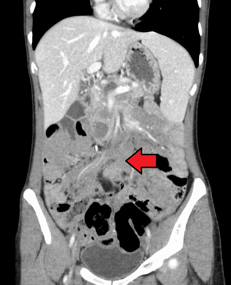
Desmoid Tumor

A desmoid tumor (DT) is a benign, locally invasive soft tissue tumor associated with a high recurrence rate but with no metastatic potential.
Epidemiology
DTs account for < 3% of soft tissue tumors. Their annual incidence is estimated to range between 1/250,000-1/500,000. They predominantly affect women and can occur between the ages of 15-60 years, but frequently during early adolescence and with a peak age of about 30 years.
Clinical description
In principle, DTs can occur in any part of the body: extra-abdominally (neck, shoulders, upper limbs, gluteal region), abdominally (originating from muscle fascia or the abdominal/chest wall), and more rarely intra-abdominally in the mesentery or retroperitoneum. Usually, they are firm and smooth palpable masses upon discovery. Depending on the location of the tumor, symptoms may include pain, fever, and functional impairment or loss of function of the organ involved. DTs may appear after surgical resections, typically after caesarian section. Intra-abdominal DTs are often observed in patients with an association of familial adenomatous polyposis (FAP) or Gardner syndrome (see these terms).
Etiology
DTs result from the proliferation of well-differentiated myofibroblasts. The exact etiopathogenetic mechanism is still unknown, but they seem to have a multi-factorial origin with hormonal and genetic factors being involved. Somatic mutations in the CTNNB1 gene (3q21) encoding beta-catenin have been found in about 85 % of sporadic cases. In cases with FAP, DTs have been associated with mutations in the tumor suppressor gene APC (5q21-q22) encoding the adenomatous polyposis coli protein.
Diagnostic methods
Initial diagnosis is based on imaging techniques (computed tomography and magnetic resonance imaging) revealing the presence of an infiltrative growing mass. Diagnosis is confirmed by tumor biopsy showing abundant collagen surrounding elongated spindle-shaped cells containing small and regular nuclei and pale cytoplasm. Immunohistological examination shows expression of muscle cell markers (e.g. actin, desmin, vimentine) and absence of CD34. Moreover, diagnosis can be confirmed by screening for mutations of CTNNB1.
Differential diagnosis
The differential diagnosis is broad with fibrosarcomas on the one extreme and myofibroblastic processes such as nodular fasciitis and even hypertrophic scars and keloids on the other. The differential diagnosis of intra-abdominal DTs includes gastrointestinal stromal tumors, solitary fibrous tumors, inflammatory myofibroblastic tumors, sclerosing mesenteritis and retroperitoneal fibrosis (see these terms).
Genetic counseling
Most cases are sporadic. Familial cases (5-10 %) are associated with FAP.
Management and treatment
Complete surgical resection remains the therapeutic mainstay of DTs. For unresectable tumors or those not amenable to surgical resection with R0 (microscopic tumor clearance) intent or accompanied by an unacceptable function loss, non-surgical treatments comprise radiotherapy, anti-estrogen therapy, non-steroidal anti-inflammatory agents, chemotherapy (e.g. methotrexate, vinblastine/vinorelbine, pegylated liposomal doxorubicin) and/or tyrosine kinase inhibitors (e.g. imatinib, sorafenib). As DTs have a variable and often unpredictable clinical course, a period of watchful waiting is advisable for asymptomatic patients. As DTs often recur, a surveillance strategy every 3-6 months is essential.
Prognosis
Local recurrence occurs in around 70 % of cases. Prognosis depends on the type of tumor. Life expectancy is normal for abdominal and extra-abdominal tumors. However, it is lower in cases of intra-abdominal DTs due to complications such as intestinal obstruction, hydronephrosis or sepsis. Repeated surgical resections are associated with a greater risk of morbidity.