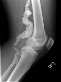
Calcification Of Joints And Arteries

A number sign (#) is used with this entry because of evidence that calcification of joints and arteries (CALJA) is caused by homozygous or compound heterozygous mutation in the NT5E gene (129190) on chromosome 6q14.
DescriptionAdult-onset calcification of the lower extremity arteries, including the iliac, femoral, and tibial arteries, and hand and foot capsule joints is an autosomal recessive condition that represents only the second mendelian disorder of isolated calcification (see generalized arterial calcification of infancy (GACI), 208000). Age of onset has been reported as early as the second decade of life, usually involving intense joint pain or calcification in the hands (St. Hilaire et al., 2011).
Clinical FeaturesSharp (1954) described a family in which 2 of 4 sibs from a first-cousin marriage displayed calcification of joint structures and arteries of an unusual type. The remaining 2 sibs and the son and daughter of one of the severely affected sibs seemed to show a milder form of the disorder affecting only arteries. Two previously reported sporadic cases were noted. Ball (1978) stated that at autopsy in 1967, one of the severely affected sibs, F.W., a 62-year-old male, showed widespread calcification of the media of the aorta and of the iliac, femoral and tibial arteries, with bilateral gangrene of the feet; calcification of the mitral and aortic valve rings and membranous portion of the interventricular septum; ossification of intervertebral discs and interspinous ligaments; calcific deposits in the capsule of metacarpophalangeal joints and lateral ligaments of the knees; and excessive bone formation in attachment of quadriceps tendon to the patella. Thus, there was evidence of both dystrophic calcification and ectopic ossification.
St. Hilaire et al. (2011) studied 3 families with calcification of the lower extremity arteries and hand and foot joints. In the first family, there were 5 affected sibs, born of unaffected third-cousin parents of English descent. The proband was a 54-year-old woman with a 20 year history of intermittent claudication and markedly reduced ankle/brachial blood pressure indices. Contrast-enhanced magnetic resonance angiography revealed extensive occlusion of the iliac, femoropopliteal, and tibial arteries, with extensive collateralization. Plain radiography of the lower extremities revealed heavy calcification with areas of arteriomegaly; chest x-ray showed no vascular calcifications above the diaphragm. Radiography also revealed juxtaarticular joint-capsule calcifications of the fingers, wrists, ankles, and feet. All 4 of her sibs had disabling intermittent claudication and similar calcifications on radiography, and 1 sister also had radiographs showing vascular and periarticular calcifications in the shoulders, elbows, hands, hips, and knees; notably absent were calcifications in the coronary arteries, except in 1 brother who had moderate coronary calcifications. Four of the sibs had joint pain and swelling; past evaluations for rheumatoid arthritis and other joint-related autoimmune disorders were negative. The second family consisted of 3 affected Italian sisters whose unaffected parents were distantly related. The proband was a 68-year-old woman who had a history of joint pain in the hands unresponsive to corticosteroids, in whom radiographs of the lower limbs revealed calcifications. Her 70- and 73-year-old sisters had lower extremity pain and similar calcifications. The proband of the third family was a 44-year-old woman with an English father and French mother, in whom dystrophic calcifications were noted in wrist films at 26 years of age; at age 41 years, swelling and severe pain in the hands and wrists were diagnosed as pseudogout. She developed mild paresthesias of the lower extremities at 42 years of age, and evaluation revealed extensive calcifications of the distal arteries, with sparing of the carotid and coronary arteries and the aorta. None of the 9 affected patients and none of their parents or children had abnormal bone morphologic characteristics, type 2 diabetes mellitus, or decreased renal function.
MappingIn a consanguineous family of English descent in which 5 sibs were affected with calcification of the lower extremity arteries and hand and foot joints, St. Hilaire et al. (2011) performed homozygosity mapping and identified a single 22.4-Mb region of homozygosity on chromosome 6q14, for which they obtained a lod score of 4.81 by parametric multipoint linkage analysis.
Molecular GeneticsIn 5 affected sibs from a consanguineous family of English descent with calcifications of arteries and joints mapping to chromosome 6q14, St. Hilaire et al. (2011) analyzed 3 candidate genes and identified homozygosity for a nonsense mutation in the NT5E gene (S221X; 129190.0001). Analysis of NT5E in 3 affected Italian sisters revealed homozygosity for a missense mutation (C358Y; 129190.0002), and the proband from a third family was found to be a compound heterozygote for S221X and a frameshift mutation (129190.0003). The mutations segregated with disease in all 3 families, and none was present in 400 alleles from ethnically matched controls.