Piebald Trait

A number sign (#) is used with this entry because piebaldism can be caused by heterozygous mutation in the KIT protooncogene (164920) on chromosome 4q12.
There is also evidence that piebaldism can be caused by heterozygous mutation in the gene encoding the zinc finger transcription factor SNAI2 (602150) on chromosome 8q11.
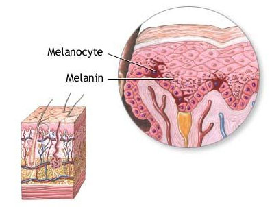
DescriptionPiebaldism is a rare autosomal dominant trait characterized by the congenital absence of melanocytes in affected areas of the skin and hair. A white forelock of hair, often triangular in shape, may be the only manifestation, or both the hair and the underlying forehead may be involved. The eyebrows and eyelashes may be affected. Irregularly shaped white patches may be observed on the face, trunk, and extremities, usually in a symmetrical distribution. Typically, islands of hyperpigmentation are present within and at the border of depigmented areas (summary by Thomas et al., 2004).
Clinical FeaturesSundfor (1939) described a family in which many persons had a white forelock, often with unpigmented patches on the forehead, limbs, and other areas of the body.
Loewenthal (1959) assigned the name albinoidism to a dominantly inherited condition characterized by a white 'blaze' in the scalp hair, usually the forelock, and/or patches of leukoderma. Epitheliomas occurred with increased frequency. The designation albinoidism is better reserved for the recessive condition simulating true albinism.
Comings and Odland (1966) found the trait in 6 generations. A genetic defect in melanoblast differentiation was postulated.
The statement that deafness does not occur in persons with the piebald trait as a pleiotropic effect of the gene may not be true. Reed et al. (1967) noted profound deafness with piebaldism in 2 patients. Some of the patients of Comings and Odland (1966) were deaf.
Winship et al. (1991) described 7 affected persons in 3 generations. Two other affected individuals were deceased. The disorder seemed distinct from Waardenburg syndrome (193500). White forelock and patches of leukoderma occur also in Waardenburg syndrome and in Fanconi anemia (227650).
From India, Mahakrishnan and Srinivasan (1980) reported Hirschsprung disease in 2 brothers who had piebaldness (white forelock, patches of depigmentation over the upper third of the forearms and the lower part of the arms, diffuse hypopigmentation of the abdomen and chest, and heterochromia iridis); their father had a white forelock also.
Hulten et al. (1987) reported a presumed homozygote; the severely affected child was born to heterozygous parents. He had complete absence of hair and pigmentation and had blue irides.
Richards et al. (2001) described a mother and her 8-year-old daughter with a phenotype of typical piebaldism but with progressive depigmentation.
InheritancePiebaldism is an autosomal dominant disorder (Thomas et al., 2004).
Keeler (1934) described a Louisiana black family in which piebaldism could be traced back to a woman born in 1853.
Selmanowitz et al. (1977) published a pedigree with at least 10 affected persons in 4 generations.
Farag et al. (1992) described a Bedouin kindred with 19 affected persons in 5 generations.
MappingLyon (1988) pointed out that the location of the W locus on mouse chromosome 5 supports the location of a piebald trait gene on chromosome 4 of man since there is a large, conserved synteny group on those chromosomes of the 2 species. Geissler et al. (1988) identified cloned DNA markers near the W locus and determined the genetic distance from a number of other loci.
A piebaldism locus in man was mapped to chromosome 4q by the identification of causative mutations in the KIT gene (Giebel and Spritz, 1991).
CytogeneticsFunderburk and Crandall (1974) reported a 3-year-old boy with moderate mental retardation, short stature, and integumentary pigment changes typical of the autosomal dominant piebald syndrome. The patient's chromosomes showed a reciprocal translocation and an intercalary deletion of one chromosome 4. Lacassie et al. (1977) found a similar case that illustrated the association of piebald trait with interstitial deletion of the long arm of chromosome 4 (4q13). The deleted segment was adjacent to centromeric heterochromatin, raising the question of position effect.
Hoo et al. (1986) described a case of de novo deletion in 4q and pointed out that several of the patients with comparable deletions have had abnormal skin pigmentation compatible with the piebald trait. Further analysis suggested that the piebald trait locus may be situated in band 4q12.
Yamamoto et al. (1989) reported piebald trait in a child with de novo interstitial deletion of 4q, specifically 4q12-q21.1. Other features included mental and motor retardation despite normal somatic growth, aplasia cutis of the scalp, flat nasal root and tip, micrognathia, widely spaced nipples, and agenesis of the right kidney.
In a patient with piebaldism, mental retardation, and multiple congenital anomalies associated with a 46,XY,del(4)(q12q21.1) karyotype, Spritz et al. (1992) identified deletion of both the KIT and the PDGFRA (173490) genes. The patient was hemizygous for the 2 deleted genes.
Molecular GeneticsIn a patient with classic autosomal dominant piebaldism, Giebel and Spritz (1991) identified heterozygosity for a missense mutation in the KIT gene (G664R; 164920.0001) that was not found in 40 controls. Genetic linkage analysis of the mutation in the proband's family, which could trace its inheritance for 15 generations, yielded a lod score of 6.02 at theta = 0.0.
Fleischman et al. (1991) analyzed the KIT gene in 7 unrelated patients with piebaldism and identified heterozygous deletion of KIT in 1 patient (164920.0002).
In affected individuals from 3 unrelated families with piebaldism, Spritz et al. (1992) identified heterozygosity for a missense mutation (F584L; 164920.0003) and 2 frameshift mutations (164920.0004; 164920.0005), respectively, in the KIT gene.
In 1 of 10 unrelated individuals with piebaldism, Fleischman (1992) identified a missense mutation in the KIT gene (E583K; 164920.0006).
In affected individuals from 2 large families segregating autosomal dominant piebaldism, Spritz et al. (1992) identified heterozygosity for a frameshift mutation (164920.0007) and a splice site mutation (164920.0008), respectively.
In a South African girl of Xhosa ancestry who had severe piebaldism and profound congenital sensorineural deafness, Spritz and Beighton (1998) identified a heterozygous missense mutation in the KIT gene (R796G; 164920.0016). Her mother and brother were reported to be similarly affected, but were not available for study.
In a Japanese mother and daughter with piebaldism, Nomura et al. (1998) identified heterozygosity for a missense mutation in the KIT gene (T847P; 164920.0019).
In affected individuals from 2 families and a sporadic patient with piebaldism, Syrris et al. (2000) identified 3 different missense mutations in the KIT gene (see, e.g., 164920.0022).
In a mother and daughter with progressive piebaldism, including total hair depigmentation in the mother, Richards et al. (2001) identified heterozygosity for a missense mutation in the KIT gene (V620A; 164920.0025). The mutation was not found in family members with a localized patch of white hair without depigmentation, or in 52 controls.
In a mother and her 8-year-old daughter, both of whom had a phenotype of typical piebaldism but with progressive depigmentation, including total hair pigmentation in the mother, Richards et al. (2001) identified heterozygosity for a novel mutation in the intracellular tyrosine kinase domain of the KIT gene (V630A; 164920.0025). The authors speculated that the mutation may cause melanocyte instability, leading to progressive loss of pigmentation as well as the progressive appearance of hyperpigmented macules.
SNAI2 Mutations
In 3 of 17 unrelated patients with piebaldism who had no 'apparent' mutations in the KIT protooncogene, Sanchez-Martin et al. (2003) identified a heterozygous deletion of the SNAI2 gene (602150.0002). Two of the patients were sporadic cases and the other had 2 affected sibs and an affected daughter; all parents were nonconsanguineous and unaffected.
Genotype/Phenotype CorrelationsThe severity of the clinical phenotype in patients with piebaldism correlates with the site of the mutation with the KIT gene. The most severe mutations tend to be dominant-negative missense mutations involving the intracellular tyrosine kinase domain. Mutations leading to an intermediate severity phenotype have largely been located at or near the transmembrane region, regardless of whether they are missense, nonsense, or frameshift mutations. Mutations leading to the mildest phenotype occur in the N-terminal extracellular ligand-binding domain with resultant haploinsufficiency (summary by Richards et al., 2001).
Animal ModelIn mice, aganglionic megacolon is associated with the piebald trait (Bielschowsky and Schofield, 1962), inherited probably as an autosomal recessive.
HistoryGeorge Catlin (1796-1872), painter of the American Indians, painted an affected Mandan Indian. Multiple members of the group were said to have been affected.