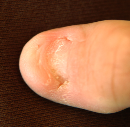
Nail-Patella Syndrome

A number sign (#) is used with this entry because of evidence that nail-patella syndrome (NPS) is caused by heterozygous mutation in the LIM-homeodomain protein LMX1B (602575) on chromosome 9q33.
Clinical FeaturesDysplasia of the nails and absent or hypoplastic patellae are the cardinal features but others are iliac horns, abnormality of the elbows interfering with pronation and supination, and in some cases nephropathy. Nephropathy was an associated abnormality in the family of Hawkins and Smith (1950). The renal change resembles glomerulonephritis. It is relatively benign although fatality at a young age from this complication has been described (Leahy, 1966). The renal disorder in the case of Simila et al. (1970) took the appearance of congenital nephrosis; 8 persons in the family had nail-patella syndrome, of whom 5 also had renal disease. The seeming familial aggregation of the renal complications suggested 2 separate genes, one for a nephropathic form and one for a nonnephropathic form. They might be allelic since no heterogeneity has been detected in the linkage with the ABO locus. As demonstrated by electron microscopy by Morita et al. (1973), among others, many collagen fibrils are present in the thickened basement membranes and in mesangial matrix of otherwise normal glomeruli. Abnormalities of collagen at this site have also been demonstrated in Alport syndrome (104200). Both of these conditions may be special forms of heritable disorders of connective tissue.
Gilula and Kantor (1975) found colon cancer in association with the nail-patella syndrome.
Sabnis et al. (1980) reported 3 patients with collagenation of glomerular basement membrane like that of the nail-patella syndrome. However, typical bone and nail changes were said to be absent. It is not clear how thoroughly the changes were sought or whether minor changes were present. An 8-year-old boy, son of first-cousin Palestinian Arabs, presented with the nephrotic syndrome. A sister and brother had died of renal disease at ages 6.5 and 9 years, respectively. A 13-year-old girl presented with recurrent urinary tract infections, proteinuria, and edema. No family information was provided. During evaluation of aortic regurgitation, a 27-year-old asymptomatic woman was noted to have proteinuria and renal insufficiency. A sister had undergone renal transplant (diagnosis unknown) and a brother had nephritis. The father's autopsy report stated 'severe interstitial nephritis and hypernephroma.' Questions include: (1) Do these cases represent variable expression of the classic nail-patella gene? (2) Is there a genetic form of glomerular basement membrane collagenosis that lacks the bone and nail involvement of the nail-patella syndrome, and is, perhaps, in light of case 1, inherited as an autosomal recessive? The range of variability of manifestation in a large series of cases ascertained through family studies has not, to my knowledge, been determined (VAM).
The Goodpasture antigen, an autoantibody, comprises the NC1 domain of the alpha-3 chain of type IV collagen (see 120070). The occurrence of Goodpasture syndrome (233450) in a patient with NPS (Curtis et al., 1976) may be more than coincidence.
This condition is sometimes called Fong disease for the physician who discovered it (Fong, 1946) in a patient on whom he performed intravenous pyelography while investigating hypertension and albuminuria related to pregnancy. No similar abnormality had been seen by many persons who reviewed the patient's films. No comments were made concerning nail dysplasia or absence of patellae. The name of Turner (1933) is associated with the disorder because of his description of 2 extensively affected families. (The designation Turner syndrome, however, leads to confusion with the XO syndrome.) He described the absence of the patellas and the inability to extend the elbows completely, as well as nail dysplasia. He also showed an x-ray of the pelvis which demonstrated the iliac horns clearly, but he made no reference to this finding in either the legend to the figure or in the text.
Aschner (1934) reported that in a study on the genetics of the human skeletal system, she had found in the literature 8 families in which defect or hypoplasia of the patella was inherited through several generations with 'a hereditary defect of the thumbnails in all the affected members' of 3 of the families. Citing her mentor Julius Bauer as the authority, Aschner (1934) suggested that the association of nail dysplasia and absence of the patella was the consequence of close linkage of 2 mutant genes. She was sharply critical of the interpretation given by Oesterreicher (1930), who described affected members in 5 generations and interpreted the association as representing 'polyphenia' (which we would call pleiotropy, or pleiotropism). Aschner (1934) wrote: 'It is rather curious, therefore, that even Oesterreicher, who is well acquainted with the related literature, accepts the hypothesis of polyphenia. In my opinion this may be understood by the fact that this author, a neurologist, did not feel sufficiently secure in genetics to form an opinion of his own in regard to the genotypic connections of the observed symptoms. He therefore sought the advice of Paula Hertwig, the biologist. Hertwig, on the other hand, was naturally not familiar enough with the clinical facts, which would make a polyphenia impossible.' Both iliac horns and renal dysplasia were clearly identified as features of this syndrome by Hawkins and Smith (1950). The deformity of the pelvis characteristic of trisomy 8 suggests somewhat the presence of iliac horns but the appearance is quite different from that in the nail-patella syndrome (Giedion, 1990).
Taguchi et al. (1988) demonstrated that characteristic ultrastructural changes in the glomerulus can be present even in patients without apparent clinical renal involvement. They reported a patient observed for 3 years, between ages 8 and 11. In a patient who had proteinuria before the age of 2 years, Browning et al. (1988) found characteristic changes in the glomeruli at age 27 months. Strong immunofluorescent staining, particularly for IgM, raised the possibility of superimposed immune complex disease. Richieri-Costa (1991) reported on 4 affected persons in a 3-generation Brazilian family with NPS. Three affected individuals had antecubital pterygium, 2 had proteinuria, and 1 had cleft lip and palate. Rizzo et al. (1993) described an Italian family in which bilateral antecubital pterygia, labeled arthrogryposis, was the presenting sign. Renal involvement was also severe in this family. From study of a large family with 30 patients with NPS (which the authors referred to as HOOD, hereditary osteoonychodysplasia), Looij et al. (1988) concluded that a person with NPS has a risk of about 1 in 4 of having a child with NPS nephropathy and a risk of about 1 in 10 of having a child in whom renal failure will develop. (It should be noted that they calculated their risk figures as a percentage in the article, i.e., 24% for the first risk, but stated the risk as a ratio, i.e., 1:4 in the abstract; these are not the same.) In the kidneys of an 18-week spontaneously aborted fetus of a mother with NPS, Drut et al. (1992) found changes which they suggested might be the basis of prenatal diagnosis by intrauterine kidney biopsy.
Sweeney et al. (2003) reviewed the phenotype of NPS by comparing the results of their study of 123 British patients with previously published studies. They suggested that neurologic and vasomotor symptoms are also part of the NPS phenotype. They found reports of neurologic symptoms in 28 of 110 patients: intermittent episodes of numbness and tingling and sometimes burning sensations in the hands and sometimes the feet, with no obvious precipitant. Distribution was in a 'glove and stocking' pattern. A 6% prevalence of epilepsy was found in the NPS study population, as contrasted with a lifetime prevalence of epilepsy in the United Kingdom of 0.4%. The neurologic symptoms were considered particularly interesting in light of the role of Lmx1b in neuronal migration in the mouse and in the developing brain. Vasomotor problems consisted of poor peripheral circulation, with very cold hands and feet, even in warm weather, and in some patients a specific diagnosis of Raynaud phenomenon. The prevalences of glaucoma and ocular hypertension were 9.6% and 7.2%, respectively. Lester sign, which consists of a zone of darker pigmentation of roughly cloverleaf or flower shape around the central part of the iris that is most pronounced in blue eyes, was observed in 54% of the patients in this study, usually bilaterally. Lester sign was no more frequent among those with glaucoma or ocular hypertension than in those without. Sweeney et al. (2003) stated that it is widely accepted that the iris configuration characteristic of Lester sign is not pathognomonic of NPS and may be seen in the general population, although at considerably lower frequency. The stellate iris that is commonly seen in Williams syndrome (194050) also is seen in the general population, but at a much lower frequency.
To examine bone mass and the prevalence of fragility fractures in patients with nail-patella syndrome, Towers et al. (2005) assessed bone mineral density (BMD) of the spine and hip in 31 adults and 12 children with mutation-confirmed NPS and 60 healthy age- and gender-matched adult controls. For the adults with NPS, BMD was 11 to 20% lower at the hip sites (P less than or equal to 0.001) and 8% lower at the spine (P less than 0.05) than that of controls. Towers et al. (2005) concluded that adults with NPS have a BMD that is 8 to 20% lower than controls, which is associated with an increase in the prevalence of fractures and scoliosis.
CytogeneticsGhiggeri et al. (1993) described a patient with an unbalanced translocation resulting in monosomy of 9q32-qter. The patient had dysplastic nails but normal patellas. She was found to have heterozygous deletion of COL5A1 (120215) and underexpression of alpha-1 chains of type V collagen by fibroblasts. Ghiggeri et al. (1993) suggested that mutations in the COL5A1 gene may be involved in the pathogenesis of the nail-patella syndrome. The patient also had skin and bone manifestations resembling those of Goltz syndrome (305600), an X-linked disorder. The patient had previously been described by Zuffardi et al. (1989), who used the deletion to map the AK1 and ORM1 (138600) loci.
Duba et al. (1998) identified a balanced t(9;17)(q34.1;q25) associated with NPS. By fluorescence in situ hybridization with probes from 9q, they narrowed the breakpoint region to a 17-cM interval between D9S262 and ABL (189980). The translocation was thought to have resulted from a break within or near the NPS gene, causing defective expression. Duba et al. (1998) suggested that the translocation may aid in the identification of the gene.
MappingThe nail-patella locus and the ABO blood group locus (110300) are linked (Renwick and Lawler, 1955). The recombination fraction is about 10% but is higher in females than in males (Renwick and Schulze, 1965). Ferguson-Smith et al. (1976) assigned the ABO-NPS1-AK1 linkage group to 9q34 by regional assignment of AK1 (103000) in studies of a chromosome deletion.
By linkage analysis in 3 families, using highly informative dinucleotide repeat polymorphisms on 9q33-q34, Campeau et al. (1995) confirmed the assignment of NPS1 to that region and localized the gene to an interval on 9q34.1 distal to D9S60 and proximal to the argininosuccinate synthetase gene (ASS; 603470).
McIntosh et al. (1997) further reduced the NPS1 candidate interval to 1-2 cM. They used 13 polymorphic markers in 5 families to position NPS1 between D9S60 and AK1. Maximum LOD scores of 27.0 and 22.0 were obtained with D9S112 and D9S315, respectively, at zero recombination. The order cen--D9S60--(D9S112/D9S315/NPS1)--AK1--tel was determined by linkage analysis.
Molecular GeneticsRenwick (1956) found evidence that the expression of this autosomal dominant disorder, nail-patella syndrome, is modified by variation in the alleles on the 'normal' chromosome. The variants of the normal allele are known as isoalleles. Evidence that they affect in trans the expression of the mutant allele on the other chromosome comes from the fact that the correlation in characteristics of the disorder, e.g., severity by some measure, between sibs is greater than the correlation between parents and offspring. Using the nail defect and the patellar size as indices of severity, Renwick (1956) found parent-child correlations of approximately zero and sib-sib correlations of approximately one-half.
Stern (1960) quoted Lionel Penrose who pointed out the likely operation of isoalleles in myotonic dystrophy (Penrose, 1948): the affected parent might be homozygous for an isoallele that favored reduced expression of the heterozygous mutation; let us symbolize the genotype d1d1. All affected children would have the d1 gene in cis with the mutation and the other chromosome carry the D1 allele making for increased expression of the mutation. Since all affected children might be heterozygous for the modifier change, the correlation between sibs should be much higher than that between affected parent and affected offspring.
Dunston et al. (2005) found no support for a role of the wildtype allele in modifying the phenotype of the nail-patella syndrome. Earlier work by Renwick (1956), subsequently expanded by Renwick and Izatt (1965), identified a correlation between affected sibs of approximately 0.50 for both nail and patellar dysplasia, in the absence of a correlation between the 2 aspects of the phenotype, or between affected parent-child pairs. The results were as expected if the modifier(s) is an allele at the NPS locus, and inherited from the unaffected parent, i.e., as an isoallele. On the other hand, Dunston et al. (2005) demonstrated association between the haplotype of the mutant allele and the variability in the nail score (p = 0.024).
The LIM-homeodomain protein Lmx1b plays a central role in dorsal/ventral patterning of the vertebrate limb. Targeted disruption of Lmx1b results in skeletal defects, including hypoplastic nails, absent patellae, and a unique form of renal dysplasia (Chen et al., 1998). Dreyer et al. (1998) showed that the LMX1B gene maps to 9q in the same region as the NPS locus by fluorescence in situ hybridization. Furthermore, they demonstrated that 3 unrelated NPS patients carried de novo heterozygous mutations in this gene. Functional studies showed that one of these mutations disrupted sequence-specific DNA binding, while the other 2 mutations resulted in premature termination of translation. These were the first described mutations in a LIM-homeodomain protein that accounted for an inherited form of abnormal skeletal patterning and renal failure.
Lichter et al. (1997) and McIntosh et al. (1997) described cosegregation of primary open angle glaucoma (POAG; 137760) and NPS. In 2 families, Lichter et al. (1997) found linkage results indicating that the cosegregation was the result of a pleiotropic effect of the NPS1 gene at 9q34. In a further study of 29 additional families, 28 of them ascertained on the basis of NPS, glaucoma was present in 9 (31%). In 1 family an individual with open angle glaucoma but no nail-patella syndrome was found; there may have been another explanation for the open angle glaucoma. That the glaucoma is a genuine pleiotropic effect of the NPS1 gene is supported by the finding of anterior segment ocular abnormalities in mice with targeted disruption of the Lmx1b gene (McIntosh, 1998). Vollrath et al. (1998) demonstrated mutations in the LMX1B gene in 4 families with combined NPS and open angle glaucoma.
Using multiplex ligation-probe amplification (MLPA) analysis, Bongers et al. (2008) identified a heterozygous deletion of the entire LMX1B gene (602575.0013) in 2 unrelated patients with nail-patella syndrome. The phenotype was similar to other reported cases with point or truncating mutations. The findings confirmed that haploinsufficiency of LMX1B is the pathogenic mechanism in nail-patella syndrome.
Genotype/Phenotype CorrelationsBongers et al. (2005) performed LMX1B mutation analysis and comprehensive examinations in 106 subjects from 32 NPS families and found that individuals with an LMXB1 mutation located in the homeodomain showed significantly more frequent and higher values of proteinuria than subjects with mutations in the LIM domains. No clear genotype-phenotype association was apparent for extrarenal manifestations.
DiagnosisPrenatal Diagnosis
Feingold et al. (1998) reported the use of ultrasound in third-trimester diagnosis of NPS. While ultrasound has the advantage of being noninvasive, the usefulness of third-trimester diagnosis is limited. McIntosh et al. (1999) used 5 DNA markers flanking the LMX1B locus to demonstrate that a fetus was affected. The pregnancy was terminated at 15 weeks. Feingold et al. (1998) considered prenatal diagnosis of NPS worthwhile because of the substantial risk for kidney disease and other associated malformations. In fact, less than 10% of the patients will develop renal failure, and the overall risk of having a renal disease is probably less than 25% (Looij et al., 1988). Most NPS families inquire about prenatal diagnosis only in the hope that severity can be predicted. It is in this regard that ultrasound may be of use in detecting early signs of severe renal damage, since there is no correlation between the LMX1B mutation and the presence of kidney disease, or overall NPS severity (McIntosh et al., 1998).
Animal ModelDunston et al. (2005) studied the expression of Lmx1b during development by inserting an internal ribosomal entry site-LacZ reporter into the 3-prime untranslated region (UTR) of the endogenous murine gene. The pattern of Lmx1b expression during the development of the limb, eye, and kidney correlated with the NPS phenotype. Additional sites of expression were observed in the central nervous system. The effects of the absence of Lmx1b in the CNS were determined in Lmx1b -/- mice by histology and immunocytochemistry. Dunston et al. (2005) concluded that Lmx1b is required for the differentiation and migration of neurons within the dorsal spinal cord, and that the inability of afferent sensory neurons to migrate into the dorsal horn was entirely consistent with diminished pain responses in some of the 16 NPS patients that they examined.
HistoryMcIntosh et al. (2005) reviewed the history of the nail-patella syndrome 50 years after the demonstration by James Renwick of linkage to the ABO blood group locus, the third autosomal linkage group identified in man. To the classical clinical tetrad of involvement of the nails, knees, and elbows and presence of iliac horns, kidney disease and glaucoma had been added as recognized parts of the syndrome. The gene mutated in NPS had been identified.