Malignant Atrophic Papulosis

Clinical Features
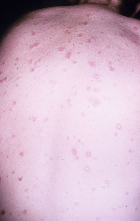
Malignant atrophic papulosis is a rare multisystem vasculopathy that affects the skin, gastrointestinal tract, and central nervous system. This condition was first described by Degos et al. (1942). Katz et al. (1997) noted that the disorder usually occurs in young adults, and affected men are said to outnumber women 3 to 1. The most common ocular manifestation is that of avascular patches on the conjunctiva, although the sclera, episclera, retina, choroid, and optic nerve may also be involved. The histologic appearance is typically characterized by a wedge-shaped area of necrosis from the epidermis through the dermis. Characteristic skin lesions are multiple asymptomatic papules with an atrophic white scarlike center surrounded by a telangiectatic border. Within weeks to years, most patients develop infarctions, mainly in the gastrointestinal and nervous systems. Systemic involvement portends a fatal outcome. There have, however, been reports of cases with only cutaneous manifestations and a favorable prognosis. The pathogenesis of Degos disease is unclear. Three main theories have been proposed: viral, autoimmune, and abnormality in coagulation.
Hall-Smith (1969) described malignant atrophic papulosis, which he referred to as a cutaneovisceral disorder, in mother and son. The 16-year-old son had a severe systemic disorder associated with profuse eruption of varioliform lesions with a sharply defined pink halo. He died 7 months after onset of the illness. At autopsy, ulcerated lesions in the intestine, similar to those seen on the skin, and associated with enlargement of the regional lymph nodes, were found. The mother was more mildly affected.
Newton and Black (1984) described the disorder in father and son. At the time of report, both showed only cutaneous manifestations of the disorder.
Katz et al. (1997) found reports of 6 families, with 6 affected members in 1 family (Kisch and Bruynzeel, 1984) and 5 in another family (Beljaards et al., 1988). Katz et al. (1997) described a family with 6 affected members distributed through 3 generations. Their proposita was a 14-year-old girl with a 1-year history of numerous asymptomatic skin lesions on her trunk and limbs. Her mother, maternal grandmother, 2 maternal great aunts, and a maternal great uncle had had similar skin lesions. The maternal great uncle had been reported by Roenigk and Farmer (1968). At age 33, the patient first reported generalized skin lesions that subsequently coalesced and reached up to 20 mm in size. Five years later he experienced bilateral pleural effusions and constrictive pericarditis. Two years later, bilateral pleural effusions recurred with subsequent gastrointestinal bleeding. During an exploratory laparotomy, diffuse infarctions similar to those in the skin were seen throughout the gastrointestinal tract. He died of gastrointestinal hemorrhage 7 years after first noting skin lesions. The patient's grandmother and mother were alive and well at the time of the Katz et al. (1997) report, with only skin lesions that originated in young adulthood. The maternal grandmother had normal screening endoscopic and colonoscopic examinations, as well as a normal evaluation of coagulation.