Coats Disease

Clinical Features
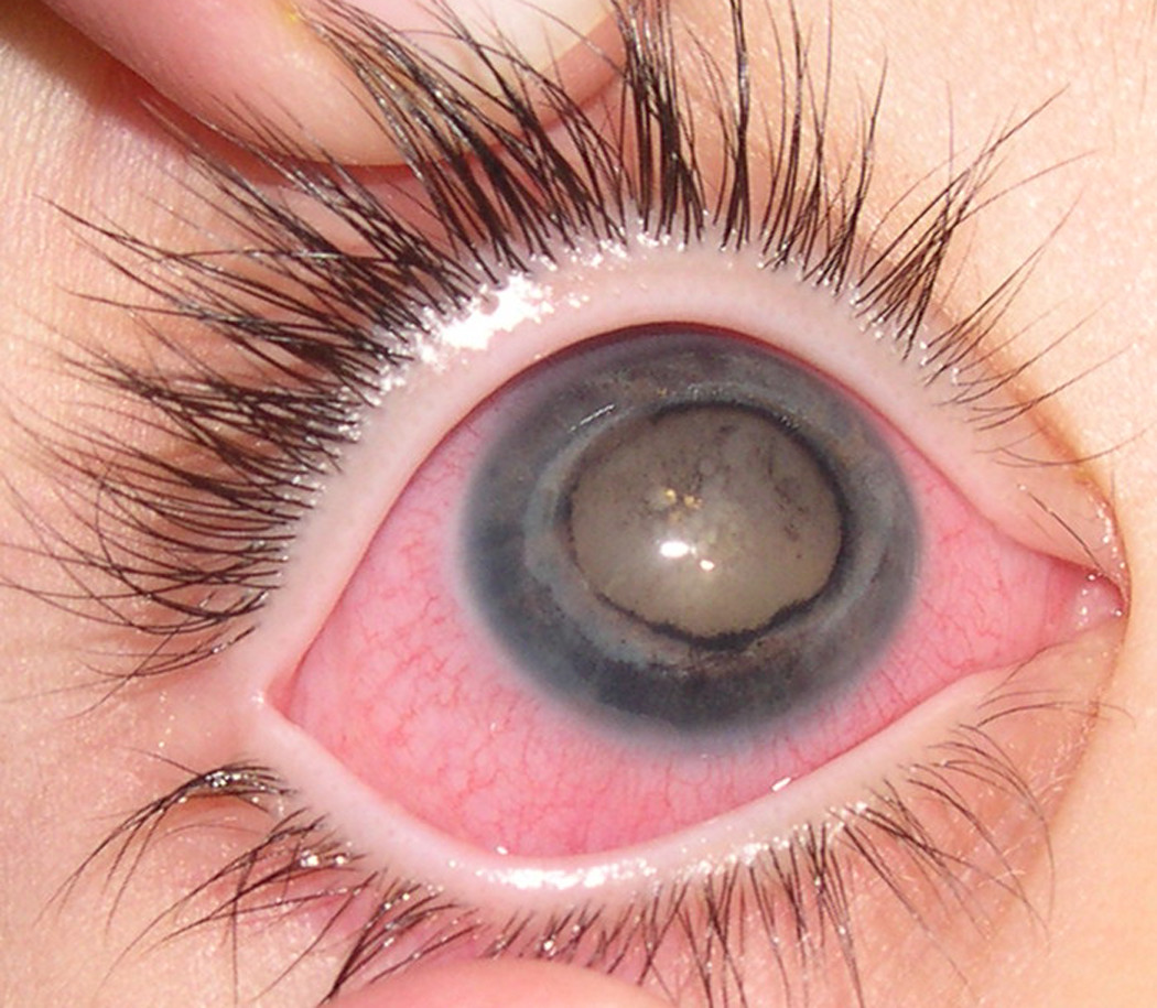
Coats disease, also called retinal telangiectasis, is a sporadic disorder characterized by a defect of retinal vascular development that results in vessel leakage, subretinal exudation, and retinal detachment. The disorder was first reported by Coats (1908) in 6 children. Initially, the condition is seen within a sector of the retina and at this stage may be associated with normal vision. However, the consequent retinal detachment often leads to progressive visual loss. In its classic form, Coats disease is almost invariably seen in males and, unlike other forms of retinal telangiectasis, is unilateral (Black et al., 1999).
In a review of reported cases of Coats disease and 150 of their own cases, Shields and Shields (2002) found that the mean age of onset of Coats disease was 5 years. Most cases were unilateral (95%) and occurred in males (75%). In most cases, the disease was slowly progressive with increasing exudative retinal detachment.
Shields et al. (2007) noted that retinal telangiectasia compatible with Coats disease can be an extramuscular manifestation of facioscapulohumeral dystrophy (FSHD; 158900) but that most affected patients have asymptomatic retinal telangiectasia found at ocular screening after diagnosis of FSHD. They described a young child who had advanced eye findings of unilateral neovascular glaucoma from bilateral retinal telangiectasia 3 years before FSHD became apparent.
Smithen et al. (2005) reported 13 patients diagnosed after the age of 35 years (mean age at diagnosis, 50 years). Those diagnosed in adulthood manifested features similar to those diagnosed in childhood: unilateral disease, male predominance, vascular telangiectasis, lipid exudation, macular edema, and areas of capillary nonperfusion with adjacent webs of filigree-like capillaries. Differences in the adult cases included limited area of involvement, slower disease progression, and hemorrhage near larger vascular dilatations.
Lim et al. (2005) described a 29-year-old woman with an unusual case of Coats disease in which there was a prominent inflammatory component in addition to the subretinal and retinal lipid crystal deposits and abnormal retinal vasculature characteristic of the disease. The patient had a 9-year history of bilateral uveitis complicated by secondary glaucoma in the right eye. Inflammation in the right eye was only partially controlled by prednisolone and cyclosporine. She developed intractable neovascular glaucoma in the left eye that led to complete loss of vision. Immunochemical staining revealed that the intraluminal inflammatory cells were macrophages and that the perivascular inflammatory infiltrates were composed largely of T lymphocytes.
Kase et al. (2013) examined the expression of vascular endothelial growth factor (VEGF; 192240) and VEGF receptor (VEGFR; 191306) in eyes enucleated because of Coats disease. Histologically, the enucleated eyes demonstrated the presence of macrophage infiltration and cholesterol clefts in the subretinal space. There were marked retinal vascular abnormalities, including dilated vessels with hyalinized vessel walls in 6 globes. Exudative retinal detachment was noted in all globes. VEGF immunoreactivity was observed in macrophages infiltrating the subretinal space and in the detached retina including several blood vessels. VEGF-positivity in macrophages was significantly higher in cases containing retinal vessel abnormalities than in those without abnormalities (P less than 0.01). VEGFR immunoreactivity was detected in endothelial cells lining the abnormal retinal vessels, where VEGFR1 (165070) and VEGFR3 (136352) were not expressed.
Molecular GeneticsBlack et al. (1999) presented evidence that Coats disease can be caused by somatic mutation in the NDP gene (300658), which is also mutant in Norrie disease (310600). They reported a woman with a unilateral variant of Coats disease who gave birth to a son affected by Norrie disease. Both carried a missense mutation within the NDP gene (cys96 to trp; 300658.0018) on Xp11.4. Subsequent analysis of the retinas of 9 enucleated eyes from males with Coats disease demonstrated in 1 a somatic mutation in the NDP gene that was not present in nonretinal tissue. This mutation was identical to that identified in the mother and son. Black et al. (1999) suggested that Coats telangiectasis is secondary to somatic mutation in the NDP gene, which results in a deficiency of norrin within the developing retina. This supported observations that the protein is critical for normal retinal vasculogenesis.