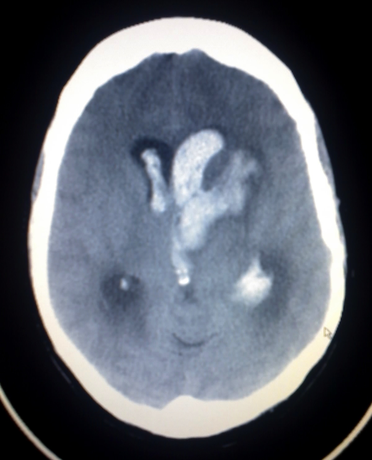
Wyburn-Mason Syndrome

Wyburn-Mason syndrome or Bonnet-Dechaume-Blanc syndrome is characterized by the association of arteriovenous malformations of the maxilla, retina, optic nerve, thalamus, hypothalamus and cerebral cortex.
Epidemiology
The prevalence is not known but it is very rare: fewer than 100 cases have been reported in the literature to date. The sex ratio is even.
Clinical description
Malformations appear successively, sometimes over several tens of years. Neurological clinical signs include: progressive neurological deficits depending on the location of the malformation, epilepsy or cephalalgia. These symptoms result in venous congestion and hemorrhage. Psychomotor delay in childhood is not common. The associated maxillofacial lesions lead to asymmetries or deformations of the face, impairment of maxillofacial osseous growth and, in intraosseous maxillo-mandibular locations, serious oral hemorrhages. The visual symptoms are caused by arteriovenous malformations of the retina and depend on the size and location of malformations (retina, optical nerve, chiasma). Partial manifestations of the syndrome (incomplete spectrum) are possible.
Etiology
Wyburn-Mason syndrome is caused by an anomaly in organogenesis, however the etiology and risk factors are unknown. There are no familial forms of the syndrome. The connection between lesions of the same angio-architectural nature, but in different locations, can be explained by the regionalized origin of cells of the vascular walls in the cephalic region, and their migration. The cells of vessels in the facial, orbital, maxillary or mandibular, and encephalic areas come from three large embryonic regions. An embryonic defect in a cellular group before its migration to its final destination may `spread' vascular lesions along the route of migration. This gives rise to cerebrofacial metameric or segmental syndromes (CAMS) called CAMS1, CAMS2 and CAMS3 according to the region from which the cells depart: CAMS1 (corpus callosum, hypothalamus, olfactory tract, forehead, nose), Wyburn-Mason syndrome that has been renamed CAMS2 (cortex and diencephalon, optic chiasma, optic nerve, retina, sphenoid, maxilla, cheek) and CAMS3 (cerebellum, temporal bone, mandible).
Diagnostic methods
Magnetic resonance imaging (MRI) is the best diagnostic instrument and provides information on the extent of the anomalies. An arteriogram then allows a more detailed analysis of the angio-architecture of the lesions, revealing the absence of capillary vessels that normally connect arteries and veins. It is most often impossible to completely treat cerebral vascular malformations because of the extent and architecture of the lesions.
Management and treatment
Targeted partial treatment through the endovascular route aims to isolate the region at risk of malformation. It can be successful in dentoalveolar and cerebral locations if a particular weakness has been identified using MRI or arteriogram. Combined management with embolization and surgery is most often necessary for maxillofacial malformations.
Prognosis
The early appearance of neurological problems is an unfavorable factor for long term prognosis.