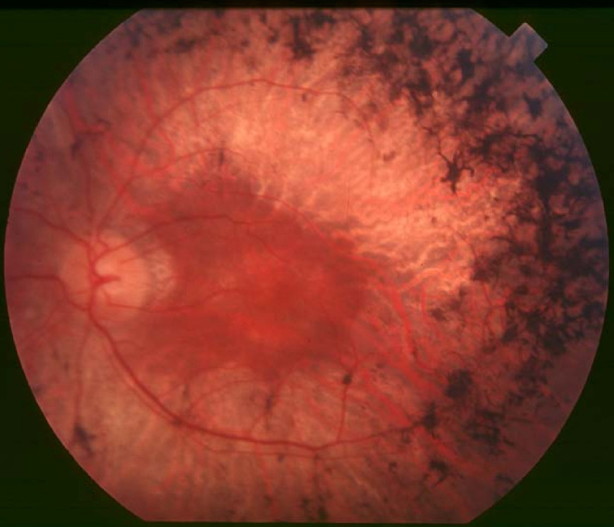
Retinopathy, Pericentral Pigmentary, Autosomal Recessive

Traboulsi et al. (1988) described a brother and sister, born to parents related as third cousins, who had pigmentary retinopathy in a pericentral distribution. The retinopathy was noted in infancy when the sibs were examined for strabismus. The optic discs, maculae, and retinal vessels were normal. Both sibs had moderate hyperopic astigmatism and esotropia. The fundus and visual acuity remained unchanged for 9 and 13 years in the brother and sister, respectively. Results of eye examinations in the father, mother, and older sister were normal. The stability of the retinal findings in visual acuity suggested a long-term favorable prognosis. Traboulsi et al. (1988) found 18 well-documented cases of pericentral pigmentary retinopathy in the literature. Although recessive inheritance had been suggested, it had never been substantiated in any of the reports. Disorders that have been labeled as central pigmentary retinopathy or inverse retinitis pigmentosa include cone-rod dystrophy (120970), Stargardt disease (248200), and Best disease (153700).
Sandberg et al. (2005) studied 18 patients, aged 32 to 65 years, with pericentral retinitis pigmentosa with follow-up for 3 to 26 years. Estimated mean annual rates of decline of remaining ocular function were 1.2% for visual acuity, 1.9% for visual field area, and 2.9% for electroretinogram amplitude for 30 Hz flashes. Sandberg et al. (2005) noted that these rates were generally slower than those previously reported for patients with typical forms of retinitis pigmentosa. Their patient sample included 2 pairs of affected sibs with normal parents and otherwise isolated cases.
See 180210 for a possible autosomal dominant form of pericentral pigmentary retinopathy.