Retinitis Pigmentosa 80

A number sign (#) is used with this entry because of evidence that retinitis pigmentosa 80 (RP80) is caused by homozygous or compound heterozygous mutation in the IFT140 gene (614620) on chromosome 16p13.
For a general phenotypic description and a discussion of genetic heterogeneity of retinitis pigmentosa, see 268000.
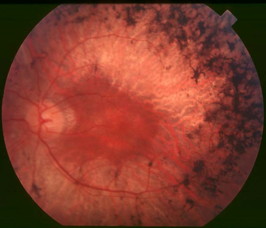
Clinical FeaturesXu et al. (2015) studied a 43-year-old Han Chinese man who developed night blindness in childhood and vision loss at age 36 years. Fundus images showed widespread retinal bone spicule pigmentation with a 'gold foil' macular reflex. Ocular coherence tomography (OCT) revealed loss of inner and outer segment photoreceptor cells. Clinical examination as well as radiography and laboratory evaluation showed no skeletal, hepatic, or renal abnormalities.
Hull et al. (2016) described 8 patients from 5 unrelated families with nonsyndromic retinitis pigmentosa and biallelic mutations in the IFT140 gene (see MOLECULAR GENETICS). Age of onset ranged from 2 years to early 30s, with night blindness or difficulty with dark and light adaptation as the presenting symptom. Fundus findings included midperipheral hypopigmentation of the retinal pigment epithelium (RPE), attenuated vessels, and macular atrophy. Electroretinography (ERG) was performed in 3 of the patients and showed markedly reduced or undetectable rod responses with subnormal cone responses, and pattern ERG was subnormal or undetectable. One of the patients was a 67-year-old British man who also had progressive hearing loss from 4 years of age, for which he wore hearing aids. Audiometry showed symmetric bilateral high-frequency hearing loss with bilateral plateau loss of 25 to 30 db in 250- to 2,000-kHz frequencies, which the authors noted was atypical for Usher syndrome (see 276900). His younger sister had RP but without hearing loss. All 8 patients had normal development without skeletal manifestations or renal failure at age 13 to 67 years.
Clinical Variability
Bifari et al. (2016) retrospectively studied 11 Saudi probands with congenital severe retinopathy, including 2 boys previously reported by Khan et al. (2014), all of whom had infantile nystagmus and poor vision since birth, with best recorded visual acuity hand motion or light perception. The fundus appeared grossly normal in children under 1 year of age, many of whom showed photophilia (staring at light). RPE changes and arteriolar attenuation were evident after age 2 years, at which time eye-rubbing (oculodigital sign) became common. In older children, peripheral mottling and sometimes peripheral punched-out chorioretinal lesions were seen. All had nonrecordable ERGs; in those who underwent further testing, autofluorescence showed increased central macular signal, and OCT showed loss of outer retinal structures. Developmental delay was observed in 7 of the probands, and the 5 probands who had x-rays of the hands all showed cone-shaped phalangeal epiphyses, with another proband exhibiting short fingers. Three of the probands (patients 4, 6, and 10a) appeared to be clinically normal other than retinal dystrophy, but did not undergo radiologic examination of the hands. The authors stated that this early childhood phenotype characterized by grossly normal fundus, high hyperopia, and nonrecordable ERG with or without neurodevelopmental delay overlapped with classic Leber congenital amaurosis (LCA; see 204000).
Molecular GeneticsIn a 43-year-old Han Chinese man with isolated retinitis pigmentosa (RP), who was negative for mutation in known RP-associated genes, Xu et al. (2015) performed whole-exome sequencing and identified compound heterozygosity for a missense mutation (L1399P; 614620.0011) and a 4-bp deletion (614620.0012) in the IFT140 gene. Screening for IFT140 variants in a collection of 243 patients with RP and 215 patients diagnosed with an early-onset form of retinal dystrophy (LCA) who were negative for mutation in known retinal disease genes revealed biallelic mutations in 6 patients, including 4 from the RP cohort and 2 from the LCA cohort (see, e.g., 614620.0013 and 614620.0014). None of the patients exhibited extraocular abnormalities.
In affected members of a large Pakistani family with nonsyndromic RP, negative for mutation in known retinal dystrophy genes, Hull et al. (2016) performed whole-exome sequencing and identified homozygosity for a T484M substitution in the IFT140 gene (614620.0013). By whole-exome sequencing in a 67-year-old Caucasian British man with RP and hearing loss, who was negative for mutation in 9 Usher syndrome-associated genes, the authors identified compound heterozygosity for a splice site mutation (614620.0002) and a missense mutation (S939P; 614620.0015) in IFT140. The proband's younger sister, who had RP without hearing loss, was also compound heterozygous for the IFT140 variants. Hull et al. (2016) also detected homozygosity for a C333Y substitution in the IFT140 gene in affected individuals from an Indian and a Pakistani family with RP, and identified a 31-year-old Caucasian British man with mild retinal changes who was compound heterozygous for a missense and a frameshift mutation in IFT140.
Bifari et al. (2016) reported 11 Saudi probands with congenital severe retinopathy, including 2 boys previously reported by Khan et al. (2014), who underwent next-generation sequencing of a panel of all retinal dystrophy-associated genes known at that time. Ten of the patients were homozygous for a missense mutation in the IFT140 gene (E664K; 614620.0001), and had no other suspicious variants in other genes on the panel. The authors suggested that E664K represented a founder mutation or a mutational hotspot. The remaining proband (patient 10a) was homozygous for a deletion/insertion mutation in IFT140 and also carried a heterozygous nonsense mutation in the ABHD12 gene (613599) and a complete deletion of the NPHP1 gene (607100).