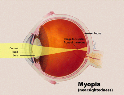
Myopia, High, With Cataract And Vitreoretinal Degeneration

A number sign (#) is used with this entry because of evidence that high myopia with cataract and vitreoretinal degeneration (MCVD) is caused by homozygous mutation in the LEPREL1 gene (P3H2; 610341) on chromosome 3q28.
Clinical FeaturesMordechai et al. (2011) studied a large consanguineous Israeli Bedouin kindred segregating autosomal recessive nonsyndromic severe myopia with variable expressivity of cataract and vitreoretinal degeneration. The 13 affected family members all presented with poor eyesight in childhood, and all had axial myopia, with increased axial lengths ranging between 25.1 mm and 30.5 mm. Eleven patients developed cataracts that were significant enough to warrant surgery in 1 or both eyes, usually in the first or second decade of life. In 3 patients, subluxated lenses were detected, and were associated with cataract in 2 patients and with lens coloboma in 1 patient. In an additional 5 patients, lens instability due to weak or partially missing lens zonules was found during cataract surgery, resulting in postoperative aphakia in 4 patients because it was impossible to insert the intraocular lens. Peripheral vitreoretinal degenerative changes were observed in 9 of the 13 affected family members, 4 of whom developed retinal tears causing retinal detachment in 1 or both eyes. In 3 patients, surgically unresponsive retinal detachments led to blindness in 1 eye. Examination by a clinical geneticist revealed no apparent dysmorphology in any of the affected individuals.
Guo et al. (2014) reported a consanguineous Chinese family in which 3 sibs had normal vision at birth but presented before 10 years of age with severe myopia (less than -12.00 diopters), followed by early-onset cataract and other ocular abnormalities. Axial lengths ranged between 28.0 mm and 32.2 mm. The proband was a male patient with severely myopic vision in both eyes, who underwent cataract extraction at 45 years of age. Funduscopy showed tigroid fundus and retinal choroid atrophy at the posterior pole. An older affected sister was blind in both eyes following cataract extraction when recruited for study. A younger sister had bilateral vision with high myopia and cataract. On funduscopy, the fundus appeared tigroid, and there was focal atrophy of choroid; in addition, the reflex of the central fovea of the macula was dull. Type B ultrasonography and optical coherence tomography also showed vitreous opacity and macular epiretinal membrane.
Khan et al. (2015) described 4 affected sisters from a consanguineous Saudi family with lens subluxation and juvenile lens opacities. The proband was a 14-year-old girl with long-standing poor vision, in whom examination revealed asymmetric bilateral temporal lens subluxation and crystalline lens opacities that were worse in the right eye. Her 22-year-old sister, who had cataract surgery at age 14 years, developed bilateral retinal breaks postoperatively that were treated with peripheral laser. At age 20, she underwent pars plana vitrectomy and scleral band placement for inferonasal rhegmatogenous detachment in the left eye. The proband's 15-year-old sister had bilateral cataract surgery at age 7 years, and had multiple bilateral rhegmatogenous retinal detachments postoperatively. The youngest sister, aged 10 years, underwent lensectomy and anterior vitrectomy for bilateral temporal lens subluxation at age 8 years. In addition, the proband had bilateral axial lengths of more than 27 mm, and 2 of her sisters had refraction values that were also indicative of long axial lengths.
MappingIn a large consanguineous Israeli Bedouin kindred segregating autosomal recessive nonsyndromic severe myopia with cataract and vitreoretinal degeneration, Mordechai et al. (2011) performed genomewide linkage analysis followed by fine mapping that indicated a minimal candidate interval of 1.7 Mb (about 4 cM) on chromosome 3q28, with a lod score of 11.5 for marker D3S1314 (theta = 0).
Molecular GeneticsIn a large consanguineous Israeli Bedouin kindred with nonsyndromic severe myopia with cataract and vitreoretinal degeneration mapping to chromosome 3q28, Mordechai et al. (2011) analyzed 6 candidate genes and identified homozygosity for a missense mutation in the LEPREL1 gene (G508V; 610341.0001) that segregated fully with disease in the family and was not found in 200 ethnically matched controls.
In 3 affected individuals from a Chinese family with high myopia and early-onset cataract with vitreoretinal degeneration, Guo et al. (2014) identified homozygosity for a nonsense mutation in the LEPREL1 gene (Q5X; 610341.0002). The mutation segregated with disease in the family and was not found in 526 Chinese controls.
In 4 affected sisters from a consanguineous Saudi family with high myopia, juvenile cataract, lens dislocation, and retinal detachment, Khan et al. (2015) sequenced the candidate gene LEPREL1 and identified homozygosity for a 1-bp deletion (610341.0003). Unaffected family members were heterozygous for the mutation. Noting that 2 of the sisters presented with lens subluxation, Khan et al. (2015) stated that LEPREL1-associated ectopia lentis is distinguishable by the presence of juvenile lens opacities and axial myopia; they also noted that there appears to be a predisposition for retinal tears/detachment following intraocular surgery.