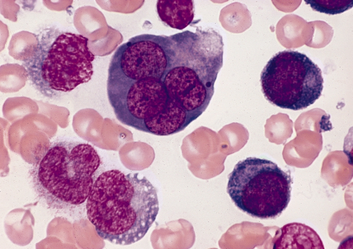
Erythroleukemia, Familial, Susceptibility To

A number sign (#) is used with this entry because of evidence that susceptibility to familial erythroleukemia (FERLK) is caused by heterozygous mutation in the ERBB3 gene (190151) on chromosome 12q13. One such family has been reported.
DescriptionFamilial erythroleukemia is a leukemic or preleukemic state in which red cell proliferation is the predominant feature. Hematologic characteristics include particularly ineffective and hyperplastic erythropoiesis with megaloblastic components accompanied by myeloblastic proliferation of varying degree (Park et al., 2002).
Park et al. (2002) discussed the evolution of the definition of 'erythroleukemia,' which is considered by most to be a subtype of acute myelogenous leukemia (AML; 601626). Controversy about the precise definition of erythroleukemia revolves around the number or percentage of erythroblasts and myeloblasts found in the bone marrow and peripheral circulation. In the French-American-British (FAB) classification system (Bennett et al., 1985), it is known as AML-M6, whereas in the revised World Health Organization (WHO) classification system (Harris et al., 1999), it is known as 'AML, not otherwise categorized' (Zini and D'Onofrio, 2004).
Clinical FeaturesDavidson et al. (1978) reported the clinical and hematologic details of an affected brother and sister. Their father presumably had died of a similar disease. Both sibs showed chromosomal changes, a finding previously reported in a majority of sporadic cases. The authors could find no 'documented evidence of the familial occurrence' of erythroleukemia.
Nissenblatt et al. (1982) diagnosed erythroleukemia in 3 brothers in a 6-month period in 1976. A son of 1 brother had died with erythroleukemia 5 years earlier. Among close relatives of these men, 14 of 16 tested had elevated IgM (mean, 352.8 mg per dl; normal, less than 145 mg per dl). This was neither a monoclonal protein nor rheumatoid factor. Erythrocyte hexokinase was elevated in 23 of 24 persons (mean, 35 units per 100 ml RBC; normal, less than 18 units). Karyotypes in 18 relatives were normal; one leukemic showed hypoploidy and a marker chromosome.
Lee et al. (1987) provided follow-up of the family reported by Nissenblatt et al. (1982), noting that there were 6 affected individuals who developed a similar disease during their sixth to eighth decades. Bone marrow of patients showed dyserythropoiesis with erythroid hyperplasia, increased blasts, and ringed sideroblasts. Some of the patients had secondary anemia, thrombocytopenia, and hepatosplenomegaly. Karyotype analysis of cancer cells showed multiple cytogenetic abnormalities. There was also a significant history of solid cancers in this family, including colon cancer, lung cancer, cervical cancer, breast cancer, and esophageal cancer.
Peterson et al. (1984) described a family in which 4 of 11 sibs were affected. A fifth sib developed unexplained marrow hypoplasia. Cytogenetic and environmental studies revealed no explanation for the familial clustering. The authors suggested that the natural history of the disorder in this sibship was initial reactive changes in the marrow with subsequent progression to myelodysplasia with sideroblastosis, and finally to Di Guglielmo syndrome. The proband, aged 55 years, was investigated because 'most of my family have leukemia' and was found to have marrow hypocellularity. The deceased members of the family, all with well-documented erythroleukemia, were a 26-year-old brother who died 14 months after clinical presentation; a 28-year-old man who died 6 months after presentation; his 39-year-old MZ twin who died 5 months after presentation; and a 41-year-old woman who died 7.5 years after mild anemia and leukopenia were discovered incidentally. The only consanguinity in the family was probably irrelevant; the paternal grandparents were distantly related.
InheritanceThe transmission pattern of familial erythroleukemia in the family reported by Braunstein et al. (2016) was consistent with autosomal dominant inheritance with incomplete penetrance.
Molecular GeneticsIn 2 living affected members of the family with familial erythroleukemia originally described by Nissenblatt et al. (1982) and Lee et al. (1987), Braunstein et al. (2016) identified a heterozygous missense mutation in the ERBB3 gene (A1337T; 190151.0002). The mutation, which was found by whole-exome sequencing, was not found in the Exome Variant Server, but was found at a low frequency in the ExAC database (7 of 119,654 alleles). The variant was also found in an asymptomatic 63-year-old family member, indicating incomplete penetrance, as well as in a female family member who died from metastatic breast cancer, but did not have leukemia. The family had a significant history of various solid tumors. Expression of the mutation into murine pro-B cells (BaF3) and human hematopoietic progenitor cells caused a growth advantage and resulted in increased cellular proliferation compared to wildtype. However, the increased proliferation was observed only in the presence of ERBB2 (164870), NRG1B (see 142445), and EPO (133170), indicating that the growth was dependent on ligand binding and heterodimerization. Analysis of the cell cycle under these conditions showed a decrease in the G1 phase and an increase in the S and G2/M phase in cells carrying the mutation compared to controls. These findings suggested that the A1337T variant overcomes a G1 phase cell cycle block. Cells carrying the mutation also showed a block in erythroid differentiation compared to controls.
Associations Pending Confirmation
Le Couedic et al. (1996) identified a heterozygous mutation in the EPOR gene (133171.0003) in 1 of 10 cases of erythroleukemia. However, in that patient, Mitjavila et al. (1991) had previously shown that erythroid growth was due to autocrine stimulation by erythropoietin (EPO; 133170). Although the role of the EPOR mutation in erythroleukemia was unclear, Le Couedic et al. (1996) noted that mutations in the Epor gene had been described in experimental erythroleukemia in mice. The EPOR mutation was found in EBV-derived cell lines, suggesting that it was germline and not somatic, although no familial cases were found. Mitjavila et al. (1991) and Le Couedic et al. (1996) suggested that in rare cases, erythroleukemia may be due to abnormal EPO signaling.
HistoryBain (2003) provided a biographic sketch of Di Guglielmo (1886-1961).