Desmoid Disease, Hereditary

A number sign (#) is used with this entry because hereditary desmoid disease has been found to be caused, at least in some cases, by mutation in the APC gene (611731) on chromosome 5q22.2.
A somatic mutation in the beta-catenin gene (CTNNB1; 116806) has been observed in a desmoid tumor derived from a patient with sporadic disease.
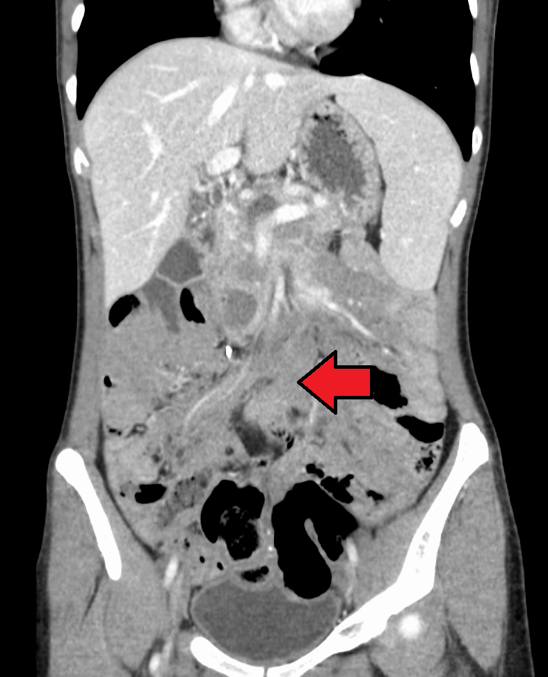
DescriptionHereditary desmoid disease usually presents as an extraintestinal manifestation of familial adenomatous polyposis (FAP; 175100), also known as Gardner syndrome, which is an autosomal dominant disorder caused by germline mutation in the APC gene. The desmoid tumors are usually intraabdominal and, although benign, can be locally aggressive and result in significant morbidity. Desmoid tumors can also arise sporadically (Couture et al., 2000).
Clinical FeaturesMaher et al. (1992) described a mother and son with marked infiltrative fibromatosis of the mesentery of the type that had been observed as part of the Gardner syndrome. However, colonic polyps, osteomas, sebaceous cysts, and congenital hypertrophy of the retinal pigment epithelium (CHRPE) typical of Gardner syndrome were not observed. In the mother, the diagnosis of infiltrative fibromatosis of the mesentery was made at age 30. Barium enema was negative for colonic polyps. She died at 32 years of age of bowel perforation and intractable abdominal sepsis; no colonic polyps were discovered postmortem. The son was found to have an abdominal mass at age 14 years. Colonoscopic examination was negative, and other extracolonic features of Gardner syndrome were not found. Family history revealed a maternal history of nonpolyposis colon cancer syndrome and breast tumor. Maher et al. (1992) noted that affected members of this family may have had a mutation at the APC locus that predisposed them to the development of desmoid tumor and perhaps colon cancer, but not multiple polyposis.
Eccles et al. (1996) described a family in which members of 3 generations showed hereditary desmoid disease characterized by multifocal fibromatosis of the paraspinal muscles, breast, occiput, arms, lower ribs, abdominal wall, and mesentery. The authors stated that some of these locations were unusual for desmoids observed in classic FAP, which usually occur in the bowel or abdominal wall. Osteomas and epidermal cysts were also observed. One patient developed carcinoma of the head of the pancreas. None of the patients had classic colonic polyposis, but 1 had a small number (less than 50) colonic polyps and 3 had fundic gland polyps. Eccles et al. (1996) proposed the name 'hereditary desmoid disease.' In an accompanying editorial, Lynch (1996) suggested that the family reported by Eccles et al. (1996) had an attenuated form of FAP with a high predilection for the development of desmoid tumors.
Couture et al. (2000) described a large French-Canadian family with hereditary desmoid disease. The phenotype was characterized by the early onset of multiple desmoid tumors arising near the axial skeleton and in proximal extremities. Although the penetrance of desmoid tumors was nearly 100%, the expression of the disease was variable among the different affected relatives. The most severely affected patient died due to persistent tumor growth in the cervical spine area, while other patients had an indolent course of disease with lesions located mainly on the proximal limbs. The proband who brought the family to attention was found to have a mass in the neck region at the age of 28 years. She subsequently developed multifocal tumors in the cervical area, occiput, arms, breast, and the thoracic paraspinal area. The initial lesions on the neck and extremities were excised, but disease relapse occurred at both local and distant sites. At the age of 46 years, the patient had good functional status with minor to moderate disability. Her son developed 3 lesions on his right upper extremity at 18 years. Her daughter had 1 lesion on the upper extremity at 16 years. Many gene carriers also had cutaneous cysts. Polyposis of the colon was rarely observed in the affected individuals, and no upper gastrointestinal polyps were documented.
Molecular GeneticsSen-Gupta et al. (1993) identified a somatic deletion in the APC gene in desmoid tissue derived from an FAP patient with a deletion at 5q.
In affected members of the family reported by Maher et al. (1992), Scott et al. (1996) identified a germline deletion in the APC gene (611731.0026). Affected members of 2 other apparently unrelated families with desmoid tumors had the same mutation, and haplotype analysis suggested a common origin. Scott et al. (1996) concluded that FAP and hereditary desmoid disease are allelic, and that APC mutations that truncate the APC protein distal to the beta-catenin-binding domain are associated with desmoid tumors, absence of congenital hypertrophy of the retinal pigment epithelium, and variable but attenuated polyposis expression.
In affected members of a family with hereditary desmoid disease, Eccles et al. (1996) identified a heterozygous germline mutation in the 3-prime end of exon 15 of the APC gene (611731.0025). There was somatic loss of the wildtype APC allele within several desmoid tumors.
Halling et al. (1999) identified a truncating mutation in the APC gene (611731.0040) in affected members of an Amish family with autosomal dominant desmoid disease.
In a large French-Canadian kindred with a hereditary desmoid disease, Couture et al. (2000) identified a heterozygous mutation in the APC gene (611731.0045). A desmoid tumor from the proband carried a somatic APC mutation (611731.0046).
Somatic Mutations
In desmoid tumor tissue from a patient with sporadic disease, Shitoh et al. (1999) identified a somatic mutation in the gene encoding beta-catenin (116806.0003). The patient had no personal or family history of FAP.
Animal ModelCyclooxygenase-2 (COX2; 600262) is an enzyme involved in prostaglandin synthesis that modulates the formation of colonic neoplasia, especially in cases due to mutations resulting in beta-catenin stabilization. Poon et al. (2001) found that human aggressive fibromatoses and lesions from mice heterozygous for the 1638N mutation of the Apc gene (a murine model for Apc-driven fibromatosis) demonstrated elevated COX2 levels. COX2 blockade, either by the selective agent DFU or by nonselective COX-blocking agents, resulted in reduced proliferation in human tumor cell cultures. Mice fed various COX-blocking agents showed a decline in tumor size. Poon et al. (2001) concluded that although COX blockade alone does not cause tumor regression, it may have a role as an adjuvant therapy to slow tumor growth in this lesion.