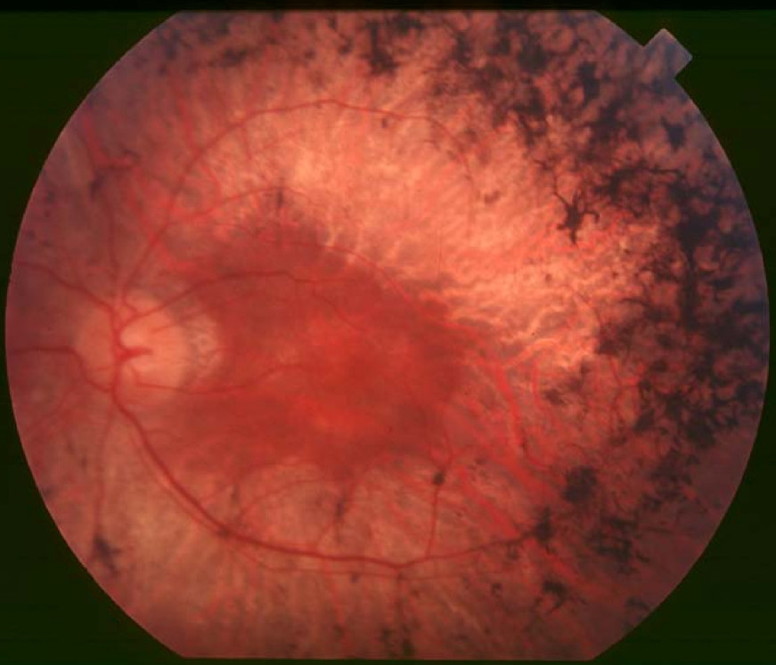
Retinitis Pigmentosa 7

A number sign (#) is used with this entry because of evidence that retinitis pigmentosa-7 (RP7) is caused by heterozygous mutation in the RDS gene (PRPH2; 179605) on chromosome 6p21.
Homozygous mutation in the PRPH2 gene causes retinal dystrophy of earlier onset, diagnosed clinically as Leber congenital amaurosis (LCA18). One patient was reported to have juvenile retinitis pigmentosa.
Heterozygous mutation in the PRPH2 gene has also been reported to cause various retinal disorders with overlapping or related phenotypes, e.g., retinitis punctata albescens (see 136880), central areolar choroidal dystrophy (CACD2; 613105), vitelliform macular dystrophy-3 (VMD3; 608161), and patterned macular dystrophy (169150).
A digenic form of retinitis pigmentosa resulting from a mutation in the RDS gene (179605.0004) and a null mutation of the ROM1 gene (see 180721.0001) has been reported.
For a phenotypic description and a discussion of genetic heterogeneity of retinitis pigmentosa, see 268000; for Leber congenital amaurosis, see 204000.
Clinical FeaturesFarrar et al. (1991) reported a large Irish family with late-onset autosomal dominant retinitis pigmentosa. The early stages of the disease were characterized by pigmentary deposits mainly in the inferior retinal midperiphery. There was loss of superior visual fields initially with subsequent retraction of the visual fields. Reduced rod amplitudes with better preservation of cone amplitudes had been detected by ERG in affected individuals from 5 years of age. Night blindness occurred in the fourth decade with loss of peripheral visual fields. Rod and cone responses were lost by the fifth or sixth decade.
Kikawa et al. (1994) identified a family with an autosomal dominant form of retinitis pigmentosa in which bull's-eye maculopathy was also present. The features were night blindness, usually noticed by the patient in the early teens; decreased visual acuity, with an onset in the late thirties; diffuse pigmentary retinal degeneration in the midperipheral to peripheral retina; and bull's-eye maculopathy, which also appeared in the late thirties. ERG assessments showed almost extinguished amplitudes of rod-isolated responses and severely reduced amplitudes of cone-isolated responses beginning at about age 9 years, even though the patient had no complaint of difficulty with night vision.
Clinical Variability
Weleber et al. (1993) reported the occurrence of 3 separate retinal phenotypes within a single family. The mother presented at age 63 with adult-onset retinitis pigmentosa that progressed dramatically over 12 years, with marked loss of peripheral visual field. One daughter developed patterned macular dystrophy (169150) at age 31 years. At age 44 years, her ERG was moderately abnormal, but her clinical disease was limited to the macula. Another daughter presented at age 42 years with macular degeneration; over 10 years, she developed a clinical picture consistent with fundus flavimaculatus. Her peripheral visual field was preserved but her ERG was moderately abnormal. A son had onset of macular degeneration at age 44 years. Pericentral scotomas were present and ERG was markedly abnormal.
Manes et al. (2015) reported the clinical findings in 27 to 67 French patients (depending on the examinations performed) with autosomal dominant RP caused by mutation in the PRPH2 gene. Severity was generally moderate, with 78% of eyes having 1.0-0.5 (20/20-20/40) visual acuity and 52% of eyes retaining more than 50% of the visual field. Some patients showed vitelliform or macular involvement. In some families, pericentral RP or macular dystrophy was found in some members, whereas widespread RP was present in other members of the same families.
Early-onset Retinal Dystrophy
Wang et al. (2013) studied 3 unrelated probands with early-onset forms of retinal dystrophy (EORD) who were homozygous for mutations in the PRPH2 gene. One proband was a 29-year-old woman who was diagnosed with Leber congenital amaurosis at 8 months of age; funduscopy showed prominent multilobulated central atrophic maculopathy surrounded by concentric rings of yellow deposits, with vessel narrowing and fine diffuse salt and pepper-like peripheral retinal changes. Her asymptomatic 57-year-old mother had 20/20 visual acuity but exhibited a florid butterfly-shaped macular pattern dystrophy (see 169150) as well as other retinal flecks on examination, and her 7-year-old son, who had decreased visual acuity due to partial amblyopia, had a miniature form of foveal butterfly-shaped macular pattern dystrophy that was consistent with an early-stage PRPH2-related phenotype. She also had an unaffected brother with normal visual acuity and no maculopathy on examination. The second proband was a 66-year-old female with onset of disease at birth who was diagnosed with LCA; funduscopy showed pigment deposits both peripherally and in the macular region, as well as extensive central atrophic maculopathy with choroidal sclerosis, vessel narrowing, optic disc pallor, and bone-spicule pigmentation. Optical coherence tomography (OCT) confirmed the extensive maculopathy and also showed unusual globular lesions in the foveal region. The third proband was a 30-year-old woman with onset of disease at 9 months of age who was diagnosed with juvenile RP, in whom funduscopy revealed diffuse retinal dystrophy with retinal vessel narrowing, retinal pigment epithelium mottling and loss, and a multilobulated foveal abnormality. OCT showed inner segment/outer segment junction confined to the central macula, consistent with her fairly good visual acuity (20/40), and unusual-appearing deposits in the foveal areas. Her asymptomatic 56-year-old father, who had normal visual acuity, exhibited macular pattern dystrophy and foveal changes on examination. All 3 probands had constricted visual fields, and electroretinography showed reduced to nondetectable responses.
MappingIn a large Irish family with autosomal dominant retinitis pigmentosa, Farrar et al. (1991) found linkage to several markers on chromosome 6p (e.g., lod score of 6.081 at D6S105). A series of overlapping multipoint analyses yielded a maximum lod score of 6.6. The authors suggested that the human equivalent of the mouse rds gene may be involved. In a follow-up of the same family, Jordan et al. (1992) demonstrated cosegregation of an intragenic marker derived from the RDS gene with late-onset autosomal dominant retinitis pigmentosa (lod = 5.46 at theta = 0.00). Using the CEPH reference panel, they did a multipoint analysis which produced a lod score of 8.21, maximizing at the PRPH2/RDS locus.
Molecular GeneticsIn a large Irish family with late-onset autosomal dominant retinitis pigmentosa, Farrar et al. (1991) identified a 3-bp deletion in the RDS gene (179605.0001). Wells et al. (1993) identified the same mutation in another family with autosomal dominant retinitis pigmentosa.
In 4 unrelated patients with RP, Kajiwara et al. (1991) identified heterozygous mutations in the RDS gene (179605.0002-179605.0004).
In a 75-year-old woman who developed progressive retinitis pigmentosa at 63 years of age, Weleber et al. (1993) identified heterozygosity for a 3-bp deletion in the RDS gene (179605.0017). She had 3 affected children with various eye phenotypes who were also heterozygous for the RDS mutation, including a 44-year-old daughter diagnosed with patterned macular dystrophy at age 31 (169150), a 49-year-old son who had onset of macular degeneration at age 44 years, and a 50-year-old daughter with progressive macular degeneration from age 42 who exhibited a clinical picture consistent with fundus flavimaculatus (see 248200).
In 3 unrelated patients with early-onset retinal dystrophy who were negative for mutation in known LCA or juvenile RP genes, Wang et al. (2013) identified homozygosity for mutations in the PRPH2 gene: 2 of the patients, 1 diagnosed with Leber congenital amaurosis (LCA) and 1 with juvenile RP, were homozygous for the L185P mutation previously detected in patients with digenic RP7 (179605.0004), whereas the third patient, diagnosed with LCA, was homozygous for another missense mutation in PRPH2 (C213R; 179605.0023).
Manes et al. (2015) screened for mutations in the PRPH2 gene in a cohort of 310 families, originating mainly from France, with autosomal dominant RP, and identified 15 different mutations in 32 probands, accounting for a prevalence of 10.3% in this population.
Digenic Mutations
In 3 unrelated families with RP, 1 of which included a patient who was previously reported by Kajiwara et al. (1991), Kajiwara et al. (1994) demonstrated that the L185P mutation (179605.0004) causes retinitis pigmentosa only when combined with a null mutation of the ROM1 gene in double heterozygous state; see 180721.0001.
NomenclatureFarrar et al. (1991) originally designated the form of RP linked to chromosome 6 as RP6, but changed the designation to RP7 in an erratum. The symbol RP6 is used for a form of RP that maps to the X chromosome; see 312612.