Retinitis Pigmentosa 79

A number sign (#) is used with this entry because of evidence that autosomal dominant retinitis pigmentosa-79 (RP79) is caused by heterozygous mutation in the HK1 gene (142600) on chromosome 10q22.
For a general phenotypic description and a discussion of genetic heterogeneity of retinitis pigmentosa, see 268000.
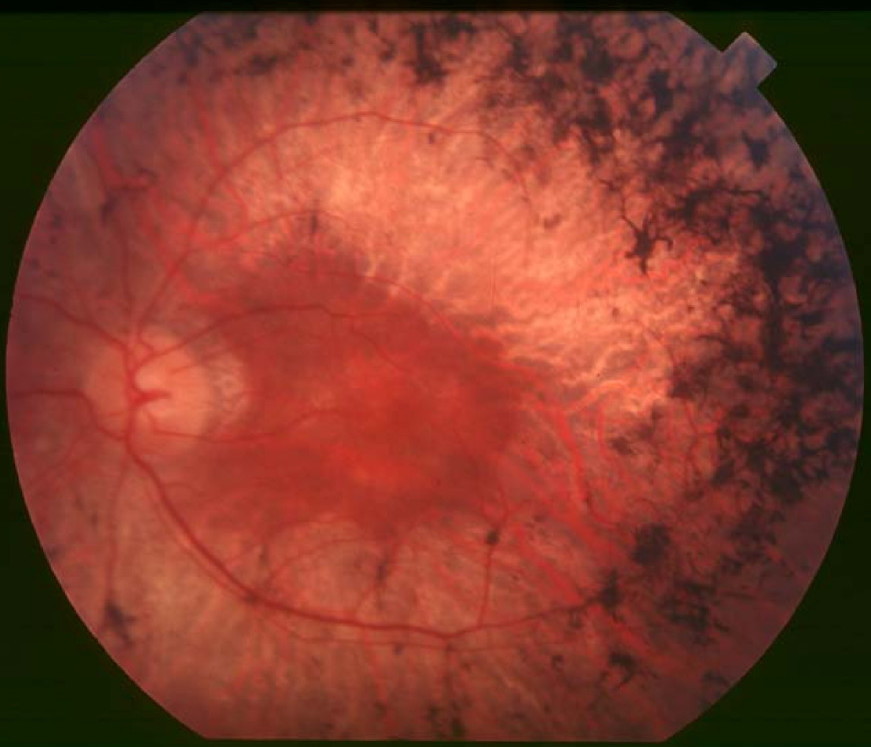
Clinical FeaturesSullivan et al. (2014) studied a large 6-generation family (UTAD003) segregating autosomal dominant retinitis pigmentosa (adRP). The retinal dystrophy in the family could be traced back to an Acadian ancestor in the early 1800s, who was from the same region of south-central Louisiana where the family still lived. Affected members of the family exhibited a highly variable phenotype, with some reporting night blindness and/or patchy vision from early childhood, whereas others did not have discernible symptoms until well into the sixth or seventh decade of life. Bone spicules, attenuated blood vessels, optic disc pallor, and peripheral atrophy were commonly seen on funduscopy. Many affected family members experienced a slow rate of vision loss and had a more pericentral pattern of degeneration and pigment deposition; 1 asymptomatic 52-year-old man showed only mild retinal changes on examination. Sullivan et al. (2014) also described patients from 4 more families with adRP, 2 from the Acadian population in Louisiana (UTAD936 and UTAD952), 1 French Canadian (MOGL1), and 1 Sicilian (MOGL2), who all carried the same HK1 mutation as patients in family UTAD003 (see MOLECULAR GENETICS). The 56-year-old female proband from family UTAD936, who was diagnosed at age 24 years, had visual fields limited to 10 degrees in all meridians, surrounded by extensive scotomas, but retained sizeable full-field electroretinographic (ERG) responses. In contrast, the 51-year-old male proband from family UTAD952, who was diagnosed as a young adult, had visual fields reduced to 30 degrees with parafoveal scotomas and undetectable responses on full-field ERGs. Both showed vessel attenuation and extensive peripheral bone spicule pigment in each eye. In family MOGL1, the father exhibited central areolar choroidal dystrophy (CACD; see 215500), whereas his 2 affected sons had pericentral RP, with striking asymmetry between the eyes. Optical coherence tomography (OCT) showed subretinal debris, with material in the inner retina. None of the affected individuals in the 5 families had extraocular manifestations, and no systemic abnormalities on glycolysis were detected.
Wang et al. (2014) reported a large 4-generation family of northern European ancestry in which 18 of 44 members had RP. Onset of reduction in night and peripheral vision ranged from age 5 to 23 years, and disease severity varied from mild pigmentary changes to severe retinal pigment epithelium (RPE) and retinal degeneration. In addition, there was concomitant cone photoreceptor degeneration in older patients, manifested by photophobia, color vision changes, and decreased central vision. Funduscopy in the proband showed typical bone spicule pigmentation and extensive retinal and RPE atrophy, as well as an abrupt border of chorioretinal degeneration outside the posterior pole, sclerosis of choroidal vessels, and pigmentary changes in the macula. Affected individuals showed no signs of anemia, exercise intolerance, or cognitive defects.
InheritanceIn the family with RP79 reported by Wang et al. (2014), the inheritance pattern was autosomal dominant with incomplete penetrance.
MappingSullivan et al. (2014) performed genomewide linkage analysis in a 6-generation family with RP and obtained a lod score greater than 3.0 in only a single 9-Mb chromosomal region at 10q21.3-q22.1. A maximum lod score of 6.2 (at theta = 0) was obtained when the HK1 mutation segregating with disease in the family (E847K; see MOLECULAR GENETICS) was analyzed as a marker at 10q22.1. Recombination events in additional families carrying the same HK1 mutation narrowed the critical interval to 55 kb between rs41279656 and D10S1742.
By genomewide linkage analysis in a large 4-generation family with RP, Wang et al. (2014) identified an 8-Mb region on chromosome 10 for which they obtained a peak parametric lod score of 3.5.
Molecular GeneticsIn a large 6-generation family with RP mapping to chromosome 10q22, in which affected members were negative for mutation in known retinal degeneration-associated genes, Sullivan et al. (2014) performed whole-exome sequencing and identified a missense mutation in the HK1 gene (E847K; 142600.0005) that segregated fully with disease and was not found in public variant databases. Analysis of the HK1 gene in 487 additional adRP probands revealed 4 more families in which the same E874K mutation segregated with disease. Haplotype analysis in the 5 families demonstrated a shared 450-kb region on chromosome 10 between markers rs41279656 and rs4746930, suggesting a founder mutation.
By whole-exome sequencing in a large 4-generation family with RP mapping to chromosome 10, in which affected members were negative for mutation in known retinal disease-associated genes, Wang et al. (2014) identified heterozygosity for the E847K mutation in the HK1 gene, which segregated with disease in the family with 85% penetrance and was not found in 11,000 in-house exomes. Two asymptomatic family members also carried the mutation; examination at ages 37 and 78 showed unaffected vision and no bone-spicule pigmentation.