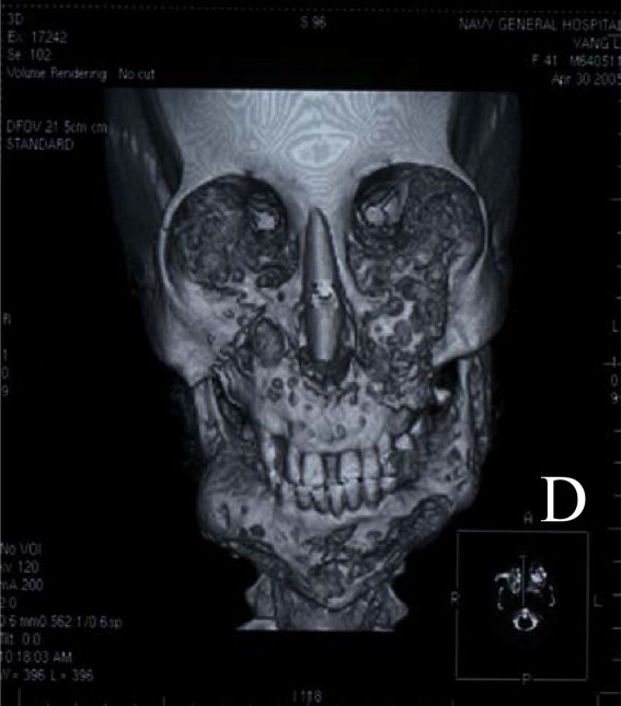
Cherubism

Cherubism is a rare, self-limiting, fibro-osseous, genetic disease of childhood and adolescence characterized by varying degrees of progressive bilateral enlargement of the mandible and/or maxilla, with clinical repercussions in severe cases.
Epidemiology
The prevalence of cherubism is unknown and is difficult to determine because of the wide clinical spectrum. About 300 cases have been reported in various ethnic groups worldwide. The disorder affects males and females equally.
Clinical description
Patients are normal at birth and most develop some degree of symmetrical mandibular and maxillary enlargement between two and five years of age. These lesions are considered aggressive, non-aggressive or quiescent depending on their clinical behavior, with each type corresponding to a particular age group (early childhood, adolescence or adulthood). In the early stages of the disease, patients may present with lymph node enlargement. The clinical manifestations are highly variable, from subclinical cases to severe enlargement causing visual, respiratory, speech, mastication, and swallowing complications. In severe cases, the fibro-osseous lesions may invade the orbital walls causing lower lid retraction, displacement of the globe, proptosis or diplopia. Respiratory complications are infrequent but can include obstructive sleep apnea, upper airway obstruction and obliteration of the nasal airway. Dental abnormalities include disrupted arrangement of primary dentition, absent teeth, rudimentary molars, premature exfoliation of primary dentition, and displacement of permanent dentition by the cystic lesions. Malocclusion is commonly found. Other organs and systems are usually not affected. Lesions progress slowly until puberty and then start to stabilize and regress through bone remodeling until the age of about 30. At this age, facial abnormalities are generally no longer visible. Cherubism can also be part of Ramon syndrome, neurofibromatosis 1 and fragile X syndrome (see these terms).
Etiology
Cherubism is caused by missense mutations in the SH3BP2 gene (4p16.3) in approximately 80% of cases, suggesting genetic heterogeneity. The exact mechanism underlying fibrous expansion has not been elucidated. Experimental data points to possible auto-inflammatory disease.
Diagnostic methods
The diagnosis is based on clinical signs, patient age, family history and radiographic findings, and can be confirmed by molecular genetic testing. Histology shows spindle cells embedded in interstitial collagen fibers and osteoclastic giant-cells.
Differential diagnosis
Differential diagnosis includes Noonan-like syndrome, hyperparathyroidism-jaw tumor syndrome, fibrous dysplasia of bone (see these terms), brown tumor of hyperparathyroidism, and central giant-cell granuloma.
Antenatal diagnosis
Prenatal diagnosis is possible when the disease-causing mutation has been detected in the family.
Genetic counseling
About 50% of cases are familial and the others appear to be related to de novo mutations. Cherubism is considered to be an autosomal dominant trait but there are reports suggesting autosomal recessive transmission. Genetic counseling is recommended for affected families.
Management and treatment
Clinical and radiographic monitoring is recommended during the growth phase of lesions. Since the disorder is usually self-limiting, surgery is not always recommended. However, surgical intervention involving resection, curettage or contouring may be required in patients with functional manifestations or for esthetic reasons and to improve quality of life. Surgery is generally indicated once lesions have become quiescent and does not alter disease progression. Careful attention should be paid to the psychological aspects related to disfigurement during childhood and adolescence.
Prognosis
Despite the possible severity of signs and symptoms, the condition is benign and the prognosis very good, with rare reports of residual deformity.