Nomenclature Other names by which Robinow syndrome has been known in the past: Costovertebral segmentation defect with mesomelia (COVESDEM): this name is no longer used because it causes confusion with similar vertebral defect syndromes, and in ROR2 -related Robinow syndrome, acromesomelia as well as mesomelia is present. Robinow-Silverman syndrome Prevalence ROR2 -related Robinow syndrome is rare. ... Vertebral anomalies and scoliosis are seen in far fewer of those with AD Robinow syndrome (<25%) compared to those with AR Robinow syndrome (>75%) [Mazzeu et al 2007]. Height is usually nearer the normal range in AD Robinow syndrome. Autosomal dominant Robinow syndrome is rarer than autosomal recessive Robinow syndrome. ... The shawl scrotum and lax ligaments of Aarskog syndrome are not found in ROR2 -related Robinow syndrome.
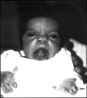
A number sign (#) is used with this entry because of evidence that autosomal dominant Robinow syndrome-2 (DRS2) is caused by heterozygous mutation in the DVL1 gene (601365) on chromosome 1p36. Description Robinow syndrome is a skeletal dysplasia characterized by distinctive facial features, including midface hypoplasia, hypertelorism, a short nose, and a broad mouth, known collectively as 'fetal facies.' ... For a discussion of genetic heterogeneity of Robinow syndrome, see RRS (268310). Clinical Features Saraiva et al. (1999) reported a pair of monozygotic twin brothers of Portuguese descent with Robinow syndrome. ... Eijkenboom et al. (2012) reported a 17-year-old girl with Robinow syndrome and low-frequency conductive hearing impairment. ... Bunn et al. (2014) reported 2 unrelated patients with Robinow syndrome. A 21-year-old woman had presented at birth with characteristic facial features, brachydactyly, camptodactyly, and clinodactyly.
For a discussion of genetic heterogeneity of Robinow syndrome, see RRS (268310). Clinical Features Robinow et al. (1969) reported a family with a short stature syndrome inherited over 6 generations. ... The authors noted similarities to the Aarskog-Scott syndrome (305400); however, the 'saddle scrotum' finding in the Aarskog-Scott syndrome may be the main differentiating feature. ... Friedman (1985) described the distinctive umbilical changes of Aarskog syndrome, Rieger syndrome (180500), and Robinow syndrome. ... Patton and Afzal (2002) compared the clinical features of the autosomal dominant and autosomal recessive forms of Robinow syndrome. The recessive Robinow syndrome tended to be more severe. ... Beiraghi et al. (2011) compared the craniofacial and intraoral phenotypes of 9 patients with dominant Robinow syndrome to 3 patients with recessive Robinow syndrome.
Robinow syndrome (RS) is a rare genetic syndrome characterized by limb shortening and abnormalities of the head, face and external genitalia. ... Clinical description Two forms of the syndrome with different patterns of inheritance and variable frequency of clinical signs have been described: a milder autosomal dominant form (autosomal dominant Robinow syndrome, see this term) and a more severe autosomal recessive form (autosomal recessive Robinow syndrome, see this term). ... Etiology Autosomal recessive Robinow syndrome is caused by mutations in the ROR2 gene (9q22). ... Differential diagnosis The main differential diagnosis is RS with a different pattern of inheritance. Syndromes that commonly involve dysmorphic facial features similar to RS, particularly hypertelorism, along with genital hypoplasia such as Aarskog-Scott syndrome and Opitz G syndrome (see these terms) should also be considered. ... Prognosis Prognosis of Robinow syndrome is generally good but the severity of heart disorders may affect life expectancy.
A number sign (#) is used with this entry because of evidence that autosomal dominant Robinow syndrome-3 (DRS3) is caused by heterozygous mutation in the DVL3 gene (601368) on chromosome 3q27. Description The clinical description of Robinow syndrome includes mesomelia, normal intellect, genital hypoplasia, and distinctive facial features comprising frontal bossing, prominent eyes, and a depressed nasal bridge, which are collectively referred to as a 'fetal face' (summary by White et al., 2016). For a discussion of genetic heterogeneity in Robinow syndrome, see RRS (268310). Molecular Genetics In a cohort of 34 individuals with a clinical diagnosis of possible Robinow syndrome, White et al. (2016) performed direct Sanger sequencing of the penultimate and final exons of the DVL1 (601365), DVL2 (602151), and DVL3 genes, and identified 1 patient with a 1-bp deletion within the final exon of DVL3 (601368.0001). Sanger sequencing the penultimate and final exons of DVL1, DVL2, and DVL3 in 17 Robinow syndrome cases from an in-house database identified 4 more patients with mutations in DVL3, including two 1-bp deletions (601368.0002-601368.0003) and 2 splice site mutations (601368.0004-601368.0005). ... White et al. (2016) stated that the phenotypic features of DVL1- and DVL3-mediated Robinow syndrome were largely concordant, but noted that only 2 of the 4 DVL3-mutated patients for whom clinical information was available had macrocephaly and all 4 had short stature, suggesting that head circumference and stature might be distinguishing features.
Robinow syndrome is a rare disorder that affects the development of many parts of the body, particularly the skeleton. ... This variant is called the osteosclerotic form of Robinow syndrome. Frequency Both the autosomal recessive and autosomal dominant forms of Robinow syndrome are rare. Fewer than 200 people with autosomal recessive Robinow syndrome have been described in the medical literature. ... Causes Autosomal recessive Robinow syndrome results from mutations in the ROR2 gene. ... Learn more about the genes associated with Robinow syndrome DVL1 DVL3 FZD2 ROR2 WNT5A Additional Information from NCBI Gene: GPC4 NXN RAC3 Inheritance Pattern As discussed above, Robinow syndrome can have either an autosomal recessive or an autosomal dominant pattern of inheritance.
The more common type of Robinow syndrome (RS) characterized by mild to moderate limb shortening and abnormalities of the head, face and external genitalia. ... In the presence of rib fusions, the recessive form of the syndrome should be considered. Etiology Mutations in WNT5A gene (3p14.3) have been reported in some patients (< 10%) with autosomal dominant Robinow syndrome.
Robinow syndrome is a rare disorder that affects the bones as well as other parts of the body. Two forms of Robinow syndrome have been described: autosomal recessive Robinow syndrome, and the milder autosomal dominant Robinow syndrome. ... This form is caused by mutations in the ROR2 gene. Autosomal dominant Robinow syndrome causes more mild, but similar, features. ... A variant type of this form is additionally characterized by osteosclerosis . Autosomal dominant Robinow syndrome may be caused by a mutation in the WNT5A or DVL1 gene. In some cases, the underlying cause of Robinow syndrome is unknown. Management may include bracing or surgery for skeletal abnormalities and growth hormone to increase growth rate in affected children.
Patients also often have polyneuropathy, liver disease, and metabolic syndrome with abnormal glucose tolerance, and hyperlipidemia. ... Other disorders to consider include Cushing syndrome, familial partial lipodystrophy, especially Dunnigan syndrome related to LMNA gene mutations, familial angiolipomatosis, Prune belly syndrome and lymphoma.
The peripheral neuropathy is often attributed to alcoholism, but the pathologic findings of Pollock et al. (1988) led them to conclude that the neuropathy is in fact an integral part of the syndrome. Biochemical observations suggested a defect in catecholamine-stimulated lipolysis at the level of cell membranes. ... Her son, who also carried the mutation, had MERRF syndrome (545000); the mother had no signs of MERRF syndrome. ... She developed dysarthria, dysphagia, and ptosis, suggestive of a stroke, and subsequently had lactic acidosis with multiorgan failure; some of these features are found in MERRF syndrome. Muscle biopsy of the proband showed both ragged-red and COX-negative fibers.
Multiple symmetric lipomatosis is a rare condition characterized by the symmetric growth of fatty tumors ( lipomas ) around the neck, shoulders, upper arms and/or upper trunk. It most often affects men of Mediterranean ancestry between the ages of 30 and 70 who have a history of alcohol abuse. Non-alcoholics and women can also be affected. The signs and symptoms vary greatly from person to person. Usually, accumulation of fatty tissue increases over time and may lead to a loss of neck mobility and pain. The lipomas can cause physical deformity and peripheral neuropathy, when they compress a nerve.
The height deficit in LWD was approximately two-thirds that of Turner syndrome. Madelung deformity was present in 74% of LWD children and adults and was more frequent and severe in females than males. The prevalence of Madelung deformity was higher in the LWD versus a Turner syndrome population. The prevalence of increased carrying angle, high-arched palate, and scoliosis was similar in the 2 populations. ... A separate comparison of 24 girls with LWD aged 1 to 15 years and 76 girls with Turner syndrome showed similar mean height deficits (SDS -2.7 for both groups). ... Overall, Madelung deformity, increased carrying angle, tibial bowing, and scoliosis were more prevalent in the LWD population, whereas high arched-palate was similarly prevalent in both LWD and Turner syndrome. Ross et al. (2005) concluded that short stature is common in both LWD girls and boys before puberty, and Turner syndrome girls. ... Ventruto et al. (1983) described a syndrome of skeletal dysplasia in 2 generations of a family.
Differential diagnosis Differential diagnoses should include the other SHOX-related haploinsufficiency disorders and related conditions such as Turner syndrome and distal monosomy Xp. Antenatal diagnosis Prenatal genetic testing is available; however, requests for testing for these disorders are uncommon but are more frequent for LMD.
Leri Weill dyschondrosteosis (LWD) is a skeletal dysplasia characterized by short stature and an abnormality of the wrist bones called Madelung deformity . Short stature is present from birth due to shortening of the long bones in the legs. Madelung deformity typically develops during mid-to-late childhood and may progress during puberty. People with this condition often experience pain in their wrists or arms. The severity of Leri Weill dyschondrosteosis varies among affected individuals, although the signs and symptoms of this condition are generally more severe in females.
A number sign (#) is used with this entry because Bart-Pumphrey syndrome is caused by heterozygous mutation in the GJB2 gene (121011) on chromosome 13q12. ... The presence of leukonychia and the absence of digital constrictions appear to distinguish this disorder from Vohwinkel syndrome (124500). A family reported by Crosby and Vidurrizaga (1976) established that keratosis palmoplantaris, probably developing only in older affected persons, is part of the syndrome. ... Palmoplantar keratoderma had locally an almost punctate surface reminiscent of Vohwinkel syndrome, and was most profound over the heels and in interphalangeal folds, resulting there in the formation of hard, hyperkeratotic bands. ... Molecular Genetics In a multigeneration Polish family with Bart-Pumphrey syndrome, Richard et al. (2004) reported a novel nonconservative missense GJB2 mutation (121011.0030) segregating with the disorder. ... In a 26-year-old male with Bart-Pumphrey syndrome, Alexandrino et al. (2005) identified heterozygosity for a missense mutation in the GJB2 gene (121011.0035).
A rare, syndromic genetic deafness disease characterized by symmetric or asymmetirc knuckle pads (typically located on the distal and interphalangeal joints), leukonychia, diffuse palmoplantar keratoderma, and congenital, mild to moderate sensorineural deafness.
Bart-Pumphrey syndrome is characterized by nail and skin abnormalities and hearing loss. People with Bart-Pumphrey syndrome typically have a white discoloration of the nails (leukonychia); the nails may also be thick and crumbly. ... The hearing loss associated with Bart-Pumphrey syndrome ranges from moderate to profound and is typically present from birth (congenital). ... Only a few affected families and individual cases have been identified. Causes Bart-Pumphrey syndrome is caused by mutations in the GJB2 gene. ... The GJB2 gene mutations that cause Bart-Pumphrey syndrome change single protein building blocks (amino acids) in the connexin 26 protein.
A number sign (#) is used with this entry because of evidence that the Rabson-Mendenhall syndrome is caused by compound heterozygous mutation in the insulin receptor gene (INSR; 147670) on chromosome 19p13. ... Biochemical Features In a 6-year-old boy with clinical features of Rabson-Mendenhall syndrome, Takata et al. (1986) found decreased insulin binding in erythrocytes, cultured fibroblasts, and transformed lymphocytes. ... In a patient with the Rabson-Mendenhall syndrome, Moncada et al. (1986) found a 90% decrease in the number of insulin receptors. ... Rittey et al. (1988) documented elevated melatonin metabolite excretion in the urine of a patient with Mendenhall syndrome presenting with neonatal hypoglycemia. ... In an English patient with Rabson-Mendenhall syndrome, Takahashi et al. (1998) identified compound heterozygosity for mutations in the INSR gene (147670.0034-147670.0035).
Severe insulin resistance in people with Rabson-Mendenhall syndrome affects the development of many parts of the body. ... Rabson-Mendenhall syndrome is one of a group of related conditions described as inherited severe insulin resistance syndromes. These disorders, which also include Donohue syndrome and type A insulin resistance syndrome, are considered part of a spectrum. Rabson-Mendenhall syndrome is intermediate in severity between Donohue syndrome (which is usually fatal before age 2) and type A insulin resistance syndrome (which is often not diagnosed until adolescence). People with Rabson-Mendenhall syndrome develop signs and symptoms early in life and live into their teens or twenties.
A rare syndrome that belongs to the group of extreme insulin-resistance syndromes (which also includes leprechaunism, the lipodystrophies, and the type A and B insulin resistance syndromes). ... Etiology As in leprechaunism (of which Rabson-Mendenhall syndrome may represent a less severe form); the condition is caused by anomalies in both alleles of the insulin-receptor gene ( INSR ;19p13.3-p13.2). ... Differential diagnosis Differential diagnoses include early-onset forms of leprechaunism, and moderate and late-onset forms of type A insulin resistance syndrome. Genetic counseling The condition is transmitted as an autosomal recessive trait and mainly affects children of consanguineous parents.
Rabson-Mendenhall syndrome (RMS) is a mild form of INSR -related severe syndromic insulin resistance, an inherited disorder associated with the inability to regulate blood sugar. ... Unregulated sugar levels over a long period of time can lead to a shortened lifespan. Donohue syndrome (leprechaunism) is a more severe form of INSR -related syndromic insulin resistance. Symptoms are similar to those seen in RMS, but are more serious. Most children with Donohue syndrome die before one year of age. Type A insulin resistance syndrome (type A) is a milder form of INSR -related syndromic insulin resistance.
A number sign (#) is used with this entry because Axenfeld-Rieger syndrome type 3 (RIEG3) is caused by heterozygous mutation in the FOXC1 gene (601090) on chromosome 6p25. For a general phenotypic description and a discussion of genetic heterogeneity of Axenfeld-Rieger syndrome, see RIEG1 (180500). See also chromosome 6pter-p24 deletion syndrome (612582), which shows phenotypic overlap with Axenfeld-Rieger syndrome type 3. ... Cunningham et al. (1998) described an autosomal dominant syndrome that combined familial Axenfeld-Rieger anomaly with atrial septal defect and sensorineural hearing loss. ... In 9 affected individuals over 3 generations of a family with Axenfeld-Rieger syndrome, Mirzayans et al. (2000) identified heterozygosity for a nonsense mutation (E23X; 601090.0005) in the FOXC1 gene. ... In a mother and son with Axenfeld-Rieger syndrome, Ito et al. (2007) analyzed the FOXC1 gene and identified a missense mutation (601090.0010) that was de novo in the mother.
Genetic Heterogeneity of Axenfeld-Rieger Syndrome Linkage studies indicate that a second type of Axenfeld-Rieger syndrome maps to chromosome 13q14 (RIEG2; 601499). ... Pearce and Kerr (1965) studied a large kindred with many affected members and emphasized the variability in expression of the syndrome. A less well-known component of this syndrome is anal stenosis (Crawford, 1967; Brailey, 1890). ... Friedman (1985) described the distinctive umbilical changes of Aarskog syndrome, Rieger syndrome, and Robinow syndrome. ... Brooks et al. (1989) suggested that this 'new' syndrome be called the short-FRAME syndrome for short stature, facial anomalies, Rieger anomaly, midline anomalies, and enamel defects. ... Vaux et al. (1992) concluded that the Rieger syndrome locus is located in either 4q25 or 4q27 inasmuch as Motegi et al. (1988) found no signs of Rieger syndrome in a patient with deletion of band 4q26.
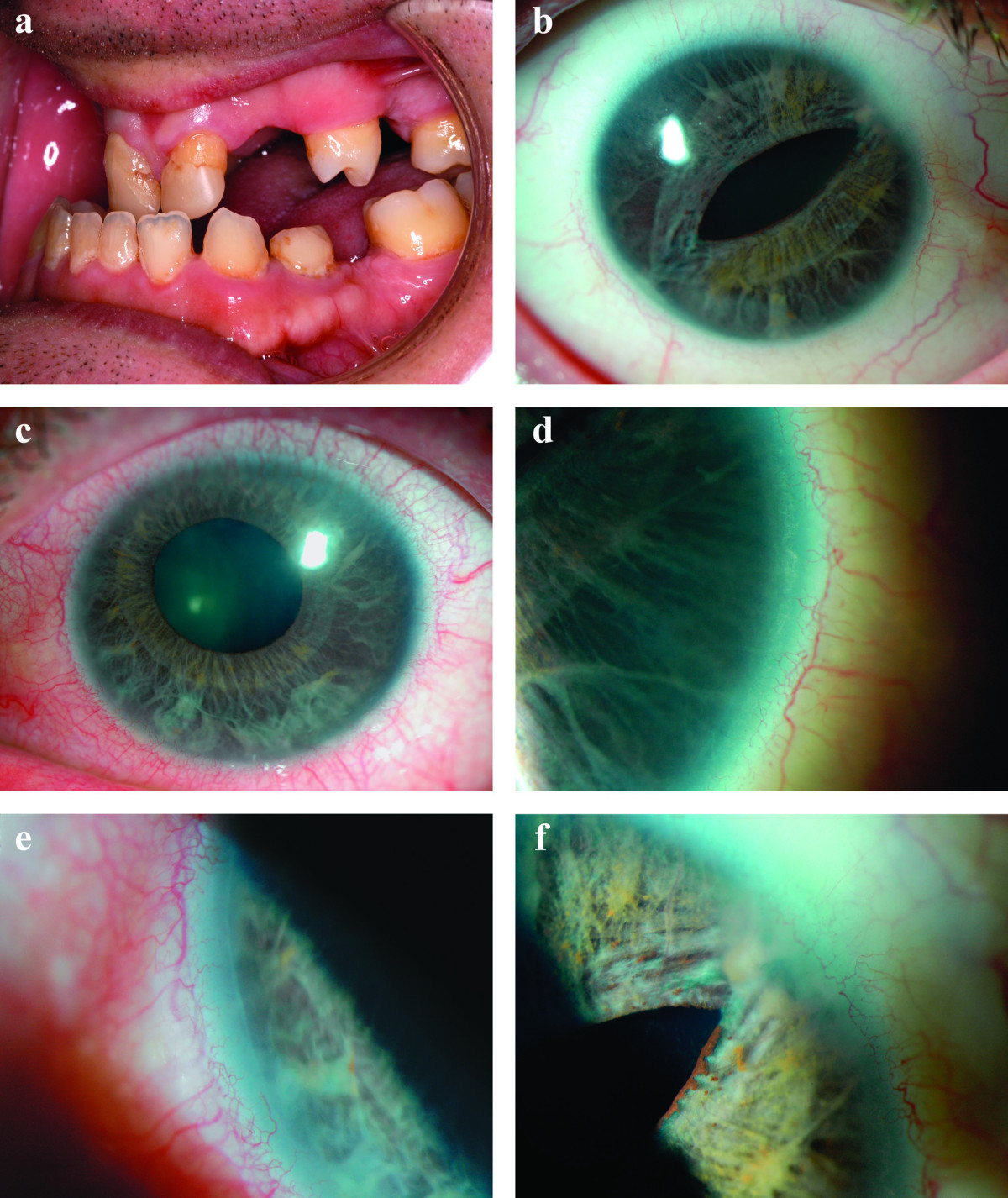
Axenfeld-Rieger syndrome (ARS) is a generic term used to designate overlapping genetic disorders, in which the major physical condition is anterior segment dysgenesis of the eye. Patients with ARS may also present with multiple variable congenital anomalies. Epidemiology The syndrome has an estimated prevalence of 1/200,000.
Axenfeld-Rieger syndrome is a group of disorders that mainly affects the development of the eye. ... Click here to view a diagram of the eye. Even though Axenfeld-Rieger syndrome is primarily an eye disorder, this syndrome can affect other parts of the body. ... Axenfeld-Rieger syndrome type 1 is caused by mutations in the PITX2 gene. Axenfeld-Rieger syndrome type 3 is caused by mutations in the FOXC1 gene. ... Axenfeld-Rieger syndrome has an autosomal dominant pattern of inheritance.
Description Axenfeld-Rieger syndrome is a disorder of morphogenesis that results in abnormal development of the anterior segment of the eye, which results in blindness from glaucoma in approximately 50% of affected individuals. ... For a general phenotypic description and a discussion of genetic heterogeneity and nomenclature of Axenfeld-Rieger syndrome, see RIEG1 (180500). Mapping Deletion of 13q14 was described in 2 cases of Rieger syndrome (Akazawa et al., 1981; Stathacopoulos et al., 1987). Phillips et al. (1996) performed linkage analysis of a large 4-generation family and demonstrated that Rieger syndrome was not linked to 4q25 but to markers on 13q14. ... The redundancy of the periumbilical skin, which has been described as a common feature of the Rieger syndrome, was found in no affected members of this kindred. ... Phillips et al. (1996) pointed to forkhead (FOXO1A; 136533) as an excellent candidate for the site of the mutation in this form of Rieger syndrome. They stated that if such mutations are found, this would be an example of a gene that can function both as an oncogene (producing rhabdomyosarcoma) and as a 'teratogene' (producing RIEG2).
Hypoparathyroidism also occurs in the autosomal dominant DiGeorge syndrome (188400) and occurs in association with candidosis and ectodermal dysplasia in the autosomal recessive syndrome of autoimmune polyendocrinopathy (240300). ... Cytogenetics Hasegawa et al. (1997) found this syndrome, which they referred to as HDR syndrome (for hypoparathyroidism, deafness, and renal dysplasia), in a Japanese girl with a de novo deletion of 10p13. The experience led them to suggest that the gene responsible for HDR syndrome is located in the 10pter-p13 region. ... None of the patients had all components of the triad of HDR syndrome, however. Van Esch et al. (1999) described 2 patients with a partial DiGeorge syndrome (facial dysmorphism, hypoparathyroidism, renal agenesis, mental retardation) and a rearrangement of chromosome 10p. ... By comparison with data previously published on patients with DiGeorge/velocardiofacial syndrome associated with 10p monosomy (see 601362), Lichtner et al. (2000) concluded that HDR is a contiguous gene syndrome.
Hypoparathyroidism-sensorineural deafness-renal disease syndrome is a rare, clinically heterogeneous genetic disorder characterized by the triad of hypoparathyroidism (H), sensorineural deafness (D) and renal disease (R). Epidemiology The exact prevalence is unknown, but the syndrome is considered to be very rare. ... Patients of both sexes have been described. Clinical description HDR syndrome may present at any age with deafness, hypocalcemia, tetany and afebrile convulsions. ... Differential diagnosis Differential diagnoses include familial idiopathic hypoparathyroidism, progressive sensorineural deafness without renal disease, autosomal recessive hypoparathyroidism with renal insufficiency and developmental delay, and deletion 22q11 syndrome. HDR syndrome should be considered in infants prenatally diagnosed with chromosome 10p defect or congenital anomalies of the kidney and urinary tract. ... Management and treatment Treatment of patients with HDR syndrome should be comprehensive and should include genetic counseling.
Barakat syndrome , also known as HDR syndrome, is a rare, genetic syndrome characterized by h ypoparathyroidism , s ensorineural deafness , and r enal (kidney) disease . ... About 65% of people with Barakat syndrome have all three of these features, while the others have various combinations of these features. Some people with Barakat syndrome have one or more of these as well as additional features. ... Hearing loss is the most consistent feature of Barakat syndrome. It is usually bilateral and can range from moderate to profound. ... For example, some people with Barakat syndrome are born with structural kidney or urinary tract abnormalities (underdeveloped or abnormally-formed), while others may have functional abnormalities (such as nephrotic syndrome , hematuria , renal tubular acidosis , or chronic kidney disease ).
Because the facial features, hypotrichosis, and anodontia seen in a patient diagnosed with Lelis syndrome (van Steensel et al., 2008) were reminiscent of hypohidrotic ectodermal dysplasia (HED; see 305100), van Steensel and van der Hout (2009) analyzed the EDA gene (300451) in this patient and identified a known causative missense mutation (R156H; 300451.0007). Van Steensel and van der Hout (2009) suggested that Lelis syndrome may be a manifestation of X-linked HED.
Möbius syndrome Other names Moebius A child with oromandibular-limb hypogenesis-Möbius syndrome. ... Limb and chest wall abnormalities sometimes occur with the syndrome. People with Möbius syndrome have normal intelligence, although their lack of facial expression is sometimes incorrectly taken to be due to dullness or unfriendliness. ... Also, because a person with Möbius syndrome cannot follow objects by moving their eyes from side to side, they turn their head instead. [ citation needed ] Culture [ edit ] Literature and media with mentions of Möbius syndrome include: [ citation needed ] The protagonist of the novel Painted by Eliza Wyatt and Christian Leffler has Möbius syndrome. ... PMID 11059047 . ^ Verzijl HT, van der Zwaag B, Cruysberg JR, Padberg GW (August 2003). "Möbius syndrome redefined: a syndrome of rhombencephalic maldevelopment". ... External links [ edit ] Classification D ICD - 10 : Q87.0 ICD - 9-CM : 352.6 OMIM : 157900 MeSH : D020331 DiseasesDB : 31978 External resources eMedicine : neuro/612 Patient UK : Möbius syndrome Möbius syndrome at Curlie Möbius syndrome at the National Institute of Neurological Disorders and Stroke v t e Congenital abnormality syndromes Craniofacial Acrocephalosyndactylia Apert syndrome Carpenter syndrome Pfeiffer syndrome Saethre–Chotzen syndrome Sakati–Nyhan–Tisdale syndrome Bonnet–Dechaume–Blanc syndrome Other Baller–Gerold syndrome Cyclopia Goldenhar syndrome Möbius syndrome Short stature 1q21.1 deletion syndrome Aarskog–Scott syndrome Cockayne syndrome Cornelia de Lange syndrome Dubowitz syndrome Noonan syndrome Robinow syndrome Silver–Russell syndrome Seckel syndrome Smith–Lemli–Opitz syndrome Snyder–Robinson syndrome Turner syndrome Limbs Adducted thumb syndrome Holt–Oram syndrome Klippel–Trénaunay–Weber syndrome Nail–patella syndrome Rubinstein–Taybi syndrome Gastrulation / mesoderm : Caudal regression syndrome Ectromelia Sirenomelia VACTERL association Overgrowth syndromes Beckwith–Wiedemann syndrome Proteus syndrome Perlman syndrome Sotos syndrome Weaver syndrome Klippel–Trénaunay–Weber syndrome Benign symmetric lipomatosis Bannayan–Riley–Ruvalcaba syndrome Neurofibromatosis type I Laurence–Moon–Bardet–Biedl Bardet–Biedl syndrome Laurence–Moon syndrome Combined/other, known locus 2 ( Feingold syndrome ) 3 ( Zimmermann–Laband syndrome ) 4 / 13 ( Fraser syndrome ) 8 ( Branchio-oto-renal syndrome , CHARGE syndrome ) 12 ( Keutel syndrome , Timothy syndrome ) 15 ( Marfan syndrome ) 19 ( Donohue syndrome ) Multiple Fryns syndrome
Verzijl et al. (2003) and Verzijl et al. (2005) concluded that HCFP and Moebius syndrome are distinct disorders, and that Moebius syndrome is a complex developmental disorder of the brainstem. Moebius syndrome was defined at the Moebius Syndrome Foundation Research Conference in 2007 as congenital, nonprogressive facial weakness with limited abduction of one or both eyes. ... Associated Syndromes Parker et al. (1981) reported at least 12 well-documented cases of association of Moebius and Poland (173800) syndromes. ... Verzijl et al. (2005) concluded that Moebius syndrome and HCFP are distinct disorders. ... Slee et al. (1991) observed deletion of 13q12.2 in a 2.5-year-old girl with Moebius syndrome. Both observations suggested that a gene responsible for Moebius syndrome is located in region 13q12.2-q13.
Moebius syndrome is a rare neurological condition that primarily affects the muscles that control facial expression and eye movement. ... Affected children often experience delayed development of motor skills (such as crawling and walking), although most eventually acquire these skills. Moebius syndrome is caused by the absence or underdevelopment of the 6th and 7th cranial nerves , which control eye movement and facial expression. Other cranial nerves may also be affected. There is no cure for Moebius syndrome, but proper care and treatment give many individuals a normal life expectancy.
Summary Clinical characteristics. Bloom syndrome (BSyn) is characterized by severe pre- and postnatal growth deficiency, immune abnormalities, sensitivity to sunlight, insulin resistance, and a high risk for many cancers that occur at an early age. ... Diagnosis Suggestive Findings Bloom syndrome (BSyn) should be suspected in an individual with any of the following clinical or cytogenetic findings. ... Option 1 When the phenotypic and laboratory findings suggest the diagnosis of Bloom syndrome molecular genetic testing approaches can include single-gene testing or use of a multigene panel : Single-gene testing. ... Clinical Characteristics Clinical Description The range of clinical features in persons with Bloom syndrome (BSyn) has been tracked through the Bloom Syndrome Registry. ... It should be recognized, however, that these recommendations are based on limited data from the Bloom Syndrome Registry and on expert opinion.
Bloom syndrome affects many different body systems and is characterized by slow growth, sun sensitivity, and an increased risk of cancer. Symptoms include short stature, sun-sensitive skin rash, and an immune system that doesn't work correctly. Some people with Bloom syndrome have learning disabilities, type 2 diabetes, and chronic obstructive pulmonary disease (COPD). Most people with Bloom syndrome develop some type of cancer by age 40. Bloom syndrome is caused by genetic variants in the BLM gene and is inherited in an autosomal recessive pattern.
Bloom syndrome is an inherited disorder characterized by short stature, a skin rash that develops after exposure to the sun, and a greatly increased risk of cancer. People with Bloom syndrome are usually smaller than 97 percent of the population in both height and weight from birth, and they rarely exceed 5 feet tall in adulthood. ... These patches appear on areas of the skin that are not exposed to the sun, and their development is not related to the rashes. People with Bloom syndrome have an increased risk of cancer. ... Causes Mutations in the BLM gene cause Bloom syndrome. The BLM gene provides instructions for making a member of a protein family called RecQ helicases. ... Genetic changes that allow cells to divide in an uncontrolled way lead to the cancers that occur in people with Bloom syndrome. Learn more about the gene associated with Bloom syndrome BLM Inheritance Pattern This condition is inherited in an autosomal recessive pattern , which means both copies of the gene in each cell have mutations.
Mori et al. (1990) reported diabetes mellitus in Bloom syndrome. Kelly (1977) observed a case of Bloom syndrome in a black female. ... (The Bloom and Fanconi syndromes are chromosome breakage or clastogenic syndromes.) ... The patient had features of both Bloom syndrome and Prader-Willi syndrome (176270). ... They also found the Bloom syndrome gene in a non-Ashkenazi Jew and reported medulloblastoma in a patient with Bloom syndrome. The hypermutability of Bloom syndrome cells includes hyperrecombinability.
Bloom syndrome is a rare disorder associated with pre- and postnatal growth deficiency, a telangiectatic erythematous rash of the face and other sun-exposed areas, insulin resistance and predisposition to early onset and recurrent cancer of multiple organ systems. Epidemiology Bloom syndrome (BSyn) overall prevalence is unknown, but in the Ashkenazi Jewish population it is estimated at approximately 1/ 48,000 births. ... Most men with BSyn have azoospermia or severe oligospermia, while women are often fertile but may begin menopause prematurely. Etiology Bloom syndrome is inherited as an autosomal recessive trait. ... Differential diagnosis The differential diagnosis of Bloom syndrome includes Fanconi anemia, Silver-Russell syndrome, Rothmund-Thomson syndrome, ataxia-telangiectasia, and Nijmegen breakage syndrome. ... Health supervision recommendations for persons with Bloom syndrome have been published, including recommendations for cancer surveillance.
Naegeli syndrome belongs to a group of disorders known as ectodermal dysplasias . This condition is characterized by absent fingerprints, thickening of the palms and soles ( palmoplantar keratoderma ), decreased sweating ( hypohidrosis ), heat intolerance, patches of darker (hyperpigmented) skin, brittle nails, abnormally colored teeth, and early tooth loss. Naegeli syndrome is caused by mutations in the KRT14 gene and inherited in an autosomal dominant manner. While there is no cure for Naegeli syndrome, treatment is based on each individual's symptoms.
A number sign (#) is used with this entry because of evidence that the Naegeli-Franceschetti-Jadassohn syndrome (NFJS) is caused by heterozygous mutation in the keratin-14 gene (KRT14; 148066) on chromosome 17q21. ... Clinical Features Naegeli (1927) described the syndrome in a father and 2 daughters. In a restudy of the original family, Franceschetti and Jadassohn (1954) documented autosomal dominant inheritance. ... NFJS and dermatopathia pigmentosa reticularis (DPR; 125595) are closely related autosomal dominant ectodermal dysplasia syndromes that clinically share complete absence of dermatoglyphics (fingerprint lines), a reticulate pattern of skin hyperpigmentation, thickening of the palms and soles (palmoplantar keratoderma), abnormal sweating, and other subtle developmental anomalies of the teeth, hair, and skin. ... Sprecher et al. (2002) studied the large Swiss family with NFJS originally described by Naegeli (1927) and assessed linkage to chromosome 17q, which was proposed to harbor the NFJ syndrome gene. Their results narrowed the NFJS locus region to 6 cM flanked by D17S933 and D17S934 with a maximum multipoint lod score of 2.7 at marker locus D17S800.
A number sign (#) is used with this entry because of evidence that Gillespie syndrome (GLSP) is caused by heterozygous mutation in the ITPR1 gene (147265) on chromosome 3p26. ... The possibility of remote consanguinity was supported by the finding of another family with this very rare syndrome in a nearby town (Sarsfield, 1971). ... Nelson et al. (1997) described 2 unrelated patients with Gillespie syndrome. The typical presentation is the discovery of fixed dilated pupils in a hypotonic infant. They considered the iris abnormality specific and pathognomonic for Gillespie syndrome. It can be distinguished clinically from other forms of aniridia and a presumptive diagnosis of Gillespie syndrome can be made in the first months of life on the basis of the ocular findings. ... The authors suggested that these findings represented a Gillespie syndrome phenotype. Donald et al. (2006) described 2 unrelated boys with Gillespie syndrome, both born of nonconsanguineous parents.
The cases referred to as atypical Gillespie syndrome correspond to those showing a more complex phenotype, associating additional ocular findings and a mild dysmorphic face. ... Differential diagnosis Differential diagnosis includes Marinesco-Sjögren syndrome (see this term) in which congenital cataract is present, as well as cerebellar ataxia, intellectual disability, and aniridia (see this term). ... Although some reported families are compatible with autosomal dominant inheritance, Gillespie syndrome is more likely to be an autosomal recessive condition.
Gillespie syndrome is characterized by underdevelopment (hypoplasia) of the colored part of the eye (the iris). ... Rapid, involuntary eye movements (nystagmus) can also occur in Gillespie syndrome. The balance and movement problems in Gillespie syndrome result from hypoplasia of the cerebellum , which is the part of the brain that coordinates movement. ... It has been estimated that Gillespie syndrome accounts for about 2 percent of cases of aniridia. Causes Gillespie syndrome is caused by mutations in the ITPR1 gene. ... However, the specific connection between these changes and the signs and symptoms of Gillespie syndrome is unclear. Learn more about the gene associated with Gillespie syndrome ITPR1 Inheritance Pattern Gillespie syndrome can be inherited in an autosomal recessive pattern , which means both copies of a gene in each cell have mutations.
Properdin deficiency was found in 9 of 46 patients in whom meningococcal disease developed after the age of 10 years. All were males. C3 deficiency syndromes (see 613799) were found in 5, and homozygous deficiency of a terminal component (C5, C6, C7, or C8) was found in 9.