• FindZebra
  • About
  • Contact
  • Help
  • Login
Advanced
  • Tk2-Related Mitochondrial Dna Maintenance Defect, Myopathic Form GeneReviews
    Clinical Manifestations of TK2 -Related Mitochondrial DNA Maintenance Defect View in own window Age of Onset Prevalence Manifestation Frequency Age <2 years (infantile onset) 61/89 (69%) Hypotonia 55/57 (96%) Elevated serum CK 56/59 (95%) Respiratory difficulties 48/53 (91%) Loss of previously acquired motor skills 43/49 (88%) mtDNA depletion 33/40 (83%) Hyporeflexia 31/39 (79%) Lactic acidemia 28/42 (67%) Motor developmental delay 18/49 (37%) Seizures 11/34 (32%) Cognitive impairment 4/43 (9%) Age 2-18 years (juvenile/ childhood onset) 14/89 (16%) Muscle weakness 8/9 (89%) mtDNA depletion 9/14 (64%) Respiratory failure 7/12 (58%) Age >18 years (adult onset) 14/89 (16%) Dysphagia 5/5 (100%) mtDNA multiple deletions 4/4 (100%) Muscle weakness 8/8 (100%) Ptosis 7/7 (100%) Ragged red fibers 8/8 (100%) Infantile onset (<2 years).
  • Scn9a Neuropathic Pain Syndromes GeneReviews
    Pathogenic missense variants in SCN9A were reported in eight of 28 adults (age ≥18 years) with inherited small fiber neuropathy (I-SFN) in whom other known causes for small fiber neuropathy (e.g., diabetes mellitus, impaired glucose tolerance, hyperlipidemia) were excluded, and who met strict diagnostic criteria for I-SFN with reduced intraepidermal nerve fiber density on skin biopsy or abnormal quantitative sensory testing [Faber et al 2012].
  • Primary Hyperoxaluria Type 3 GeneReviews
    Nephrocalcinosis of prematurity occurs in a significant proportion of infants born prior to 28 weeks' gestation and is characterized by both nephrocalcinosis and nephrolithiasis [Habbig et al 2011].
    HOGA1
  • 16p12.2 Recurrent Deletion GeneReviews
    Pizzo et al [2018] showed that among parents with the 16p12.2 deletion, 53% (17/32) had exhibited learning difficulties in school, 52% (11/21) exhibited depressive behaviors, 13% (2/15) alcohol or drug addiction, 19% (3/16) schizophrenic-like behaviors, 28% epilepsy, and 13% (2/16) bipolar disorder.
  • Glycogen Storage Disease Ia OMIM
    Obara et al. (1993) described the renal histology in 2 adult patients with type I glycogen storage disease, a 37-year-old woman and a 28-year-old man. Smit (1993) studied retrospectively 41 patients over 10 years of age from 5 different European centers.
    G6PC, HCA1, HCC, AAVS1, APOB, APOE, ARSA, SLC37A4, VWF, G6PC3
    • Glycogen Storage Disease Ic OMIM
      A number sign (#) is used with this entry because of evidence that glycogen storage disease Ic (GSD1C) is caused by homozygous or compound heterozygous mutation in the G6PT1 gene (SLC37A4; 602671), which encodes glucose-6-phosphate translocase, on chromosome 11q23. G6PT1 is also the site of the defect in glycogen storage disease Ib (GSD1B; 232220). Nordlie et al. (1983) reported studies of liver tissue from an 11-year-old girl with classic clinical features of type I glycogenosis. As in type Ib, glucose-6-phosphatase activity was lacking except in detergent-disrupted microsomes. Findings that differed from those of type Ib were interpreted on the basis of the multicomponent G6Pase system proposed by Arion et al. (1980).
    • Glycogen Storage Disease Ib OMIM
      A number sign (#) is used with this entry because of evidence that type Ib glycogen storage disease (GSD1B) is caused by homozygous or compound heterozygous mutation in the G6PT1 gene (SLC37A4; 602671), which encodes glucose-6-phosphate translocase, on chromosome 11q23. Senior and Loridan (1968) proposed the existence of a second type of von Gierke disease in which, although glucose-6-phosphatase (G6PC; 613742) activity is present on in vitro assay, glucose is not liberated from glucose-6-phosphate in vivo. They referred to this as 'functional deficiency of G6P.' They pointed out that some mutants in Neurospora show impaired enzyme function in the intact fungus despite normal activity in homogenates. Arion et al. (1975) concluded that G6Pase activity requires 2 components of the microsomal membrane: (1) a glucose-6-phosphate specific transport system that shuttles G6P from the cytoplasm to the lumen of the endoplasmic reticulum (a G6P translocase), and (2) an enzyme, glucose-6-phosphate phosphohydrolase, bound to the luminal surface of the membrane. Narisawa et al. (1978) described a patient who appeared to have a defect in the transport system.
    • Glycogen Storage Disease Type I MedlinePlus
      Glycogen storage disease type I (also known as GSDI or von Gierke disease) is an inherited disorder caused by the buildup of a complex sugar called glycogen in the body's cells. The accumulation of glycogen in certain organs and tissues, especially the liver, kidneys, and small intestines, impairs their ability to function normally. Signs and symptoms of this condition typically appear around the age of 3 or 4 months, when babies start to sleep through the night and do not eat as frequently as newborns. Affected infants may have low blood sugar (hypoglycemia), which can lead to seizures. They can also have a buildup of lactic acid in the body (lactic acidosis), high blood levels of a waste product called uric acid (hyperuricemia), and excess amounts of fats in the blood (hyperlipidemia).
    • Glycogen Storage Disease Type 1a GARD
      Glycogen storage disease type 1 is an inherited disorder caused by the buildup of a complex sugar called glycogen in the body's cells. The accumulation of glycogen in certain organs and tissues, especially the liver, kidneys, and small intestines, impairs their ability to function normally. Researchers have described two types of glycogen storage disease type 1, which differ in their signs and symptoms and genetic cause. These types are known as glycogen storage disease type IA and glycogen storage disease type IB. Glycogen storage disease type 1A is characterized by growth retardation leading to short stature and accumulation of glycogen and fat in the liver and kidneys.
  • Wolfram Syndrome 1 OMIM
    Her affected sister had died at age 28 from brainstem atrophy and central respiratory failure.
    WFS1, CISD2, GLP1R, PSPN, BPIFA2, SYVN1, NCS1, TXNIP, RIDA, NSD2, TH, STXBP3, REG1A, PSPH, ATP2A2, CAPN2, PAX3, NHS, MSMB, IL4R, HTC2, GCG, EVC, ERN1, DIAPH1, CRMP1, COMT, POU4F3
    • Wolfram Syndrome 2 OMIM
      A number sign (#) is used with this entry because of evidence that Wolfram syndrome-2 (WFS2) is caused by homozygous mutation in the CISD2 gene (611507) on chromosome 4q24. Description Wolfram syndrome-2 is an autosomal recessive neurodegenerative disorder characterized by diabetes mellitus, high frequency sensorineural hearing loss, optic atrophy or neuropathy, and defective platelet aggregation resulting in peptic ulcer bleeding (summary by Mozzillo et al., 2014). For a discussion of genetic heterogeneity of Wolfram syndrome, see WFS1 (222300). Clinical Features El-Shanti et al. (2000) reported 16 individuals from 4 consanguineous Jordanian families with a phenotype consistent with Wolfram syndrome. The patients developed diabetes mellitus in the first or second decade, followed by visual loss due to optic atrophy in all patients, and high frequency sensorineural hearing loss in all except 2 patients.
    • Wolfram Syndrome, Mitochondrial Form OMIM
      A number sign (#) is used with this entry because of evidence that a form of Wolfram syndrome may be due to mutation in mtDNA. For a general phenotypic description and a discussion of genetic heterogeneity of Wolfram syndrome, see 222300. As indicated in 222300, Rotig et al. (1993, 1993) suggested that some cases of the syndrome of early-onset diabetes mellitus, optic atrophy, and deafness may have their basis in a mitochondrial mutation. They described a patient with this disorder in association with a 7.6-kb heteroplasmic deletion of mtDNA. The deletion extended from nucleotide 6466 to nucleotide 14134, inclusive.
    • Wolfram Syndrome GARD
      Wolfram syndrome , which is also known by the acronym DIDMOAD, is an inherited condition characterized by diabetes insipidus (DI), childhood-onset diabetes mellitus (DM), a gradual loss of vision caused by optic atrophy (OA), and deafness (D). Other symptoms may include bladder and bowel dysfunction, problems with the parts of the inner ear and brain that help control balance and eye movements (vestibular deficits), temperature regulation problems, decreased balance, uncoordinated (ataxic) gait and olfactory deficits. Also, psychiatric symptoms such as anxiety and depression have also been noted in some cases. There are two types of Wolfram syndrome (type 1 and type 2) which are primarily differentiated by their genetic cause. Type 1 is caused by changes (mutations) in the WFS1 gene, while type 2 is caused by mutations in the CISD2 gene.
    • Wolfram Syndrome Orphanet
      A rare, genetic, endocrine disorder characterized by type I diabetes mellitus (DM), diabetes insipidus (DI), sensorineural deafness (D), bilateral optical atrophy (OA) and neurological signs. Epidemiology Estimated prevalence of Wolfram syndrome (WS) is 1/770,000 worldwide. Clinical description 2 types of WS may be distinguished: type 1 (WS1) and type 2 (WS2). WS1 has onset in the first decade, with DM (91% of cases) and OA (87%) manifestations. Patients present a progressive reduction of visual acuity and loss of color vision.
    • Wolfram Syndrome Wikipedia
      Human disease Wolfram syndrome Other names Diabetes insipidus-diabetes mellitus-optic atrophy-deafness syndrome Photographic image of the patient right eye showing optic atrophy without diabetic retinopathy; from Manaviat et al., 2009 [1] Specialty Medical genetics , neurology Wolfram syndrome , also called DIDMOAD ( d iabetes i nsipidus, d iabetes m ellitus, o ptic a trophy, and d eafness), is a rare autosomal-recessive genetic disorder that causes childhood-onset diabetes mellitus , optic atrophy , and deafness as well as various other possible disorders. [2] It was first described in four siblings in 1938 by Dr. Don J. Wolfram, M.D. [2] The disease affects the central nervous system (especially the brainstem ). Contents 1 Causes 1.1 WFS1 1.2 Other disorders - caused by mutations in the WFS1 gene 1.3 WFS2 2 Diagnosis 3 Treatment 4 Prognosis 5 Research 6 See also 7 References 8 External links Causes [ edit ] Wolfram syndrome was initially thought to be caused by mitochondrial dysfunction due to its symptoms and several reports of mitochondrial mutations. However, it has now been established that Wolfram syndrome is caused by endoplasmic reticulum dysfunction. [2] Two genetic forms have been described: Wolfram syndrome 1 (WFS1), [2] [3] and Wolfram syndrome 2 (WFS2). [2] [4] WFS1 [ edit ] The WFS1 or wolframin gene [5] provides instructions for making the wolframin protein. The WFS1 gene is active in cells throughout the body, with strong activity in the heart , brain , lungs , inner ear , and pancreas .
    • Wolfram Syndrome MedlinePlus
      Wolfram syndrome is a condition that affects many of the body's systems. The hallmark features of Wolfram syndrome are high blood sugar levels resulting from a shortage of the hormone insulin (diabetes mellitus ) and progressive vision loss due to degeneration of the nerves that carry information from the eyes to the brain (optic atrophy). People with Wolfram syndrome often also have pituitary gland dysfunction that results in the excretion of excessive amounts of urine (diabetes insipidus), hearing loss caused by changes in the inner ear (sensorineural deafness), urinary tract problems, reduced amounts of the sex hormone testosterone in males (hypogonadism), or neurological or psychiatric disorders. Diabetes mellitus is typically the first symptom of Wolfram syndrome, usually diagnosed around age 6. Nearly everyone with Wolfram syndrome who develops diabetes mellitus requires insulin replacement therapy.
  • Peri-Implantitis Wikipedia
    Journal of Clinical Periodontology . 28 (6): 517–523. doi : 10.1034/j.1600-051x.2001.028006517.x .
    IL1A, IL1B, TNF, TNFRSF11B, MMP8, IL6, IL1RN, OLR1, IL23A, IL22, TNFSF11, VEGFA, TLR4, TLR2, IL17A, BOP, CD14, DKK2, CPA1, CCR5, CD68, CD44, WNT5A, CD38, TNFSF12, HERPUD1, PARK7, HIF1A, CXCR3, SOST, CALR, IL21, MTG1, IL34, BRINP3, MIR27A, TNFSF12-TNFSF13, DEFB4B, TIMP2, TGFB1, TERT, SPP1, FUT4, IL1R1, FN1, IL4, FGF2, CXCL8, IL10, DEFB4A, MIP, MMP2, DEFB1, MMP9, MPO, CAD, CSF1, PPIA, CCL2, SFRP1, SNAP25, BTF3P11
  • Heterochromia Iridum Wikipedia
    Notable historical figures thought to have heterochromia include Anastasius the First , dubbed dikoros (Greek for 'having two irises'), [28] [29] and Alexander the Great , as noted by the historian Plutarch . [11] [30] In other animals [ edit ] See also: Odd-eyed cat Although infrequently seen in humans, complete heterochromia is more frequently observed in other species, where it almost always involves one blue eye. [ citation needed ] The blue eye occurs within a white spot, where melanin is absent from the skin and hair (see Leucism ).
    ACTB, PAX3, EFTUD2, ELP1, SOX10, SNAI2, PTEN, PTCH1, MITF, ACTG1, KITLG, KIT, GNAQ, GJB2, EDNRB, EDN3, ADGRV1
    • Heterochromia Iridis OMIM
      Asymmetry in the pigmentation of the irides probably occurs as an isolated phenomenon inherited as a dominant (Calhoun, 1919). Whether hereditary heterochromia iridis ever exists independent of Horner syndrome (143000), Waardenburg syndrome (193500), or the piebald trait (172800) is not clear. The melanocytes of the uveal trait constitute a branching pseudosyncytium richly innervated by sympathetic nerves. Pigmentation of the iris does not occur in the absence of this innervation. Sympathetic fibers leave the lateral horn of the gray matter of the first and second thoracic segments, pass out in the anterior roots, and join the lateral sympathetic chain via the white rami communicantes.
  • Heart Failure With Preserved Ejection Fraction Wikipedia
    (In atrial fibrillation, this additional 20% filling volume is lost and the patient may experience systolic heart failure symptoms). [28] Complete left ventricular filling is essential to maintain maximum cardiac output. ... European journal of heart failure, 13(1), 18-28. ^ González-López E, Gallego-Delgado M, Guzzo-Merello G, de Haro-Del Moral FJ, Cobo-Marcos M, Robles C, Bornstein B, Salas C, Lara-Pezzi E, Alonso-Pulpon L, Garcia-Pavia P (7 October 2015).
    REN, NR3C2, LGALS3, ST2, TTR, ALDH2, CRP, EHMT1, OPRD1, ORM1, PENK, PLG, COL18A1, TSPAN31, SI, TTN, NANS, MME, FGF23, APOL1, PDE5A, MGAM, ANKS1A, SIRT3, LAP, ALB, LEPR, EDN1, ANG, ARSL, ATP5F1A, AVP, BCHE, CYP3A4, DBP, ACE, GCG, LEP, GLP1R, CFH, NRG1, HIF1A, IGFBP7, IL1A, IL1B, IL16, ZGLP1
  • Virtual Reality Sickness Wikipedia
    Galvanic vestibular stimulation , which creates the illusion of motion by electric stimulation of the vestibular system, is another technique being explored for its potential to mitigate or eliminate the visual-vestibular mismatch. [28] Newest technology [ edit ] With the integration of virtual reality into the more commercial mainstream, issues have begun to arise in relation to VR sickness in head-mounted gaming devices. [29] While research on head-mounted VR for gaming dates back to the early 1990s, [30] the potential for mass usability has only become recently realized.
  • Sotos Syndrome 1 OMIM
    There was a strong correlation between presence of an NSD1 alteration and clinical phenotype, as 28 of 37 patients (76%) in group 1 had NSD1 mutations or deletions, whereas none of the patients in group 4 had alterations in NSD1.
    NSD1, NRK, APC2, SETD2, PIK3CA, NFIX, GPC3, STK11, EPHB2, RASIP1, FGFR3, CSPG5, FGFR4, NSD2, UROD, SET, IGF1, MAPK1, PREP, BDNF, PDC, NTF3, NGF, NFIC, ARSD
    • Sotos Syndrome GeneReviews
      Summary Clinical characteristics. Sotos syndrome is characterized by a distinctive facial appearance (broad and prominent forehead with a dolichocephalic head shape, sparse frontotemporal hair, downslanting palpebral fissures, malar flushing, long and narrow face, long chin); learning disability (early developmental delay, mild-to-severe intellectual impairment); and overgrowth (height and/or head circumference ≥2 SD above the mean). These three clinical features are considered the cardinal features of Sotos syndrome. Major features of Sotos syndrome include behavioral problems (most notably autistic spectrum disorder), advanced bone age, cardiac anomalies, cranial MRI/CT abnormalities, joint hyperlaxity with or without pes planus , maternal preeclampsia, neonatal complications, renal anomalies, scoliosis, and seizures. Diagnosis/testing. The diagnosis of Sotos syndrome is established in a proband by identification of a heterozygous NSD1 pathogenic variant or a deletion encompassing NSD1 on molecular genetic testing. Management. Treatment of manifestations: Referral to appropriate specialists for management of learning disability / speech delays, behavior problems, cardiac abnormalities, renal anomalies, scoliosis, seizures; intervention is not recommended if the brain MRI shows ventricular dilatation without raised intracranial pressure.
    • Sotos Syndrome Orphanet
      A rare genetic overgrowth syndrome characterized by a typical facial appearance, overgrowth with macrocephaly and variable intellectual impairment. Epidemiology Prevalence at birth is estimated at 1/14,000. Clinical description Excessive growth is evident across life, especially in childhood, and can manifest since fetal life., with final adult height beyond or in the upper part of the normal ranges. Macrocephaly is usually striking and disproportioned with respect to height. The distinct facial appearance is most easily recognized in early childhood (long narrow face, flushed cheeks, prominent forehead with frontotemporal hair scarcity, down-slanting palpebral fissures, hypertelorism, a high arched palate, and pointed chin). Sotos syndrome is associated with mild to severe intellectual disability as well as a wide spectrum of behavioral disorders.
    • Sotos Syndrome 3 OMIM
      A number sign (#) is used with this entry because of evidence that Sotos syndrome-3 (SOTOS3) is caused by homozygous mutation in the APC2 gene (612034) on chromosome 19p13. One such family has been reported. For a discussion of genetic heterogeneity of Sotos syndrome, see SOTOS1 (117550). Clinical Features Almuriekhi et al. (2015) reported 2 sibs, born of consanguineous Egyptian parents, with intellectual disability (IQs of 60 and 56), a severe receptive and expressive language disorder, learning disabilities, and hyperactive behavior associated with relative macrocephaly, long face, and prominent chin and nose. Brain imaging in 1 patient showed dilated lateral ventricles. Neither patient had advanced bone age, hypotonia, seizures, or autism. The findings were reminiscent of Sotos syndrome, but genetic analysis excluded mutations in the NSD1 gene (606681).
    • Sotos Syndrome GARD
      Sotos syndrome is a condition characterized mainly by distinctive facial features; overgrowth in childhood; and learning disabilities or delayed development. Facial features may include a long, narrow face; a high forehead; flushed (reddened) cheeks; a small, pointed chin; and down-slanting palpebral fissures . Affected infants and children tend to grow quickly; they are significantly taller than their siblings and peers and have a large head. Other signs and symptoms may include intellectual disability; behavioral problems; problems with speech and language; and/or weak muscle tone (hypotonia). Sotos syndrome is usually caused by a mutation in the NSD1 gene and is inherited in an autosomal dominant manner.
    • Sotos Syndrome Wikipedia
      Sotos syndrome Other names Cerebral gigantism or Sotos-Dodge syndrome Sotos syndrome is an autosomal dominant inherited condition Specialty Medical genetics Sotos syndrome is a rare genetic disorder characterized by excessive physical growth during the first years of life. Excessive growth often starts in infancy and continues into the early teen years. The disorder may be accompanied by autism , [1] mild intellectual disability , delayed motor, cognitive, and social development, hypotonia (low muscle tone), and speech impairments. Children with Sotos syndrome tend to be large at birth and are often taller, heavier, and have relatively large skulls ( macrocephaly ) than is normal for their age. Signs of the disorder, which vary among individuals, include a disproportionately large skull with a slightly protrusive forehead, large hands and feet, large mandible, hypertelorism (an abnormally increased distance between the eyes), and downslanting eyes.
  • Anomic Aphasia Wikipedia
    The studies show that verbs are harder to recall or repeat, even with rehabilitation. [24] [28] Other methods in treating anomic aphasia include circumlocution-induced naming therapy (CIN), wherein the patient uses circumlocution to assist with his or her naming rather than just being told to name the item pictured after given some sort of cue.
    GRN, MAPT, PSEN1, CHMP2B, TREM2, TMEM106B, C9orf72, APOE
  • Hyperhidrosis Wikipedia
    ] [23] [24] A microwave -based device has been tried for excessive underarm perspiration and appears to show promise. [25] Tap water iontophoresis as a treatment for palmoplantar hyperhidrosis was originally described in the 1950s. [26] Studies showed positive results and good safety with tap water iontophoresis. [27] One trial found it decreased sweating by about 80%. [28] Surgery [ edit ] Sweat gland removal or destruction is one surgical option available for axillary hyperhidrosis (excessive underarm perspiration).
    BIRC3, SCN11A, SRCAP, AGPAT2, GNA14, PLAA, CRLF1, AIP, PHOX2B, BCL10, SUCLG1, SERPINB7, CDK13, ELP1, IKBKG, CUL4B, VHL, UCP2, TP53, THPO, TERT, TERC, MLX, HNF1A, TAT, ABCC8, SPR, MALT1, KIF1B, ASCL1, CLCF1, CAVIN1, NLRP3, LONP2, GPR101, CTC1, USB1, WNK1, KLC2, CDH23, NLRC4, SLURP1, NGLY1, TMEM127, NHP2, NOP10, WRAP53, MAGEL2, RETREG1, RTEL1, SEPSECS, FOXP1, BSCL2, TINF2, PTPN22, FLRT1, SLCO2A1, SLC18A2, SLC12A3, SDHD, IGH, HPGD, HNF4A, HMBS, HLA-B, HINT1, HEXB, GDNF, GABRA3, FUCA1, FOS, FGFR3, EDN3, DKC1, DDC, CTSB, COL6A3, COL6A2, COL6A1, CLCNKB, CAV1, CACNA1S, DST, BLM, KIF1A, IGHMBP2, IL12B, JAK2, MEN1, SDHC, SDHB, SCN9A, RET, PSMB8, PRNP, MAP2K2, MAP2K1, PPARG, PARN, MPL, MAX, JUP, LIFR, KRT17, KRT16, KRT14, KRT9, KRT6B, KRT6A, KRT5, KRT1, KCNJ11, KCNA1, KCNJ18
    • Hyperhidrosis Mayo Clinic
      Overview Hyperhidrosis (hi-pur-hi-DROE-sis) is excessive sweating that's not always related to heat or exercise. You may sweat so much that it soaks through your clothes or drips off your hands. Heavy sweating can disrupt your day and cause social anxiety and embarrassment. Hyperhidrosis treatment usually helps. It often begins with antiperspirants. If these don't help, you may need to try different medications and therapies.
  • Anosmia Wikipedia
    Loss of smell has also been found to be more predictive of COVID-19 than all other symptoms, including fever, cough or fatigue, based on a survey of 2 million participants in the UK and US. [16] Google searches for "smell", "loss of smell", "anosmia", and other similar terms increased since the early months of the pandemic, and strongly correlated with increases in daily cases and deaths. [17] Research into the mechanisms underlying these symptoms are currently ongoing. [18] Many countries list anosmia as an official COVID-19 symptom, and some have developed "smell tests" as potential screening tools. [19] [20] In 2020, the Global Consortium for Chemosensory Research, a collaborative research organization of international smell and taste researchers, formed to investigate loss of smell and related chemosensory symptoms. [21] List of causes [ edit ] Upper respiratory tract infection (such as sinusitis , the common cold ) [22] COVID-19 [23] [24] Nasal polyps [25] Idiopathic hypogonadotropic hypogonadism Hypothyroidism Head trauma, damage to the ethmoid bone [26] Dementia with Lewy bodies Tumors of the frontal lobe Antibiotics Fibromyalgia Multiple sclerosis Hypoglycaemia Diabetes Asthma or allergy Hayfever Chronic obstructive pulmonary disease (COPD) Long term alcoholism Cushing's syndrome Exposure to a chemical that burns the inside of the nose Stroke Epilepsy Radiation therapy to the head and neck Liver or kidney disease Parkinson's disease [27] Alzheimer's disease [28] Toxins (especially acrylates , methacrylates [29] and cadmium [30] [31] ) Old age [32] Kallmann syndrome Primary ciliary dyskinesia Post-perfusion syndrome Laryngectomy with permanent tracheostomy Esthesioneuroblastoma is an exceedingly rare cancerous tumor that originates in or near the olfactory nerve .
    LPAR1, ADCY3, DNAI1, ARNT2, MFN2, SEMA3A, SMCHD1, ATP13A2, FLRT3, NSMF, IL17RD, HS6ST1, CHD7, WDR11, PROK2, SPRY4, KISS1R, PROKR2, CCDC141, SEMA3E, FGF17, ANK1, OTX2, ARSL, DCC, DUSP6, FGF8, FGFR1, GNRH1, ANOS1, PEX7, HESX1, PHYH, POLR2F, SCN9A, SOX2, SOX3, SOX10, TACR3, FEZF1
  • Infection Wikipedia
    A review of chronic wounds in the Journal of the American Medical Association's "Rational Clinical Examination Series" quantified the importance of increased pain as an indicator of infection. [28] The review showed that the most useful finding is an increase in the level of pain [likelihood ratio (LR) range, 11–20] makes infection much more likely, but the absence of pain (negative likelihood ratio range, 0.64–0.88) does not rule out infection (summary LR 0.64–0.88). ... Antimicrobial stewardship is the concept that healthcare providers should treat an infection with an antimicrobial that specifically works well for the target pathogen for the shortest amount of time and to only treat when there is a known or highly suspected pathogen that will respond to the medication. [47] Epidemiology [ edit ] Deaths due to infectious and parasitic diseases per million persons in 2012 28–81 82–114 115–171 172–212 213–283 284–516 517–1,193 1,194–2,476 2,477–3,954 3,955–6,812 Disability-adjusted life year for infectious and parasitic diseases per 100,000 inhabitants in 2004. [48] no data ≤250 250–500 500–1000 1000–2000 2000–3000 3000–4000 4000–5000 5000–6250 6250–12,500 12,500–25,000 25,000–50,000 ≥50,000 In 2010, about 10 million people died of infectious diseases. [49] The World Health Organization collects information on global deaths by International Classification of Disease (ICD) code categories .
    MBL2, TNF, CRP, G6PD, POMC, SAMD9, PEPD, IL18, IFNG, CXCL8, IL1B, TGFB1, IL10, TLR4, CCR5, TP53, TLR3, IRF3, APOBEC3G, TLR2, TRIM5, IL6, GHRL, BCL2, IL4, IFNA1, CD81, CXCL10, CD209, HLA-DRB1, CCL5, PTGS2, CXCL12, STAT1, PDCD1, MIF, CCL2, MX1, MAPK1, CLEC4M, CD14, CDKN2A, TLR9, PML, IFNB1, IRF1, MMP9, ACE2, EGFR, CXCR4, DEFB4A, CFTR, EIF2AK2, GZMB, DDX58, MAPK3, ICAM1, CD274, SCARB1, DAXX, IFNGR1, CAMP, IL2, BIRC5, OAS1, CD4, IL12B, IL13, TNFSF10, DEFA1, VEGFA, LTA, LEP, TNFRSF10B, CXCR3, SFTPD, CXCL5, MUC1, HLA-E, MICB, HLA-G, IDO1, SOCS3, ABL1, VDR, NFKB1, CTLA4, FAS, CD1D, CASP3, APOE, TNFRSF8, FOXP3, MAVS, CXADR, IL1RL1, CD80, TAP1, IL7, TAP2, SERPINA1, IL4R, IFNAR1, BAG3, IL1RN, PLA2G5, SSTR3, IGF2, CD38, MMP2, APOBEC3F, PON1, DEFB103B, PPIA, SP110, PRF1, IL12RB1, PCNA, IL15, APOB, MPO, TNFRSF1B, MSN, CEACAM1, BPI, EZR, FASLG, LMAN1, IFIH1, TP53AIP1, NOS2, IL16, KLRC1, CYLD, CALCA, OGG1, IRF7, IL21, CASP9, CAV1, TERT, SOD2, CD63, CD44, CDH1, AKT1, EFNB2, MAPK14, SLC11A1, GSTP1, IL22, IFNL1, PTHLH, TBK1, SLAMF1, PTPRC, PTX3, EIF4G1, KLRK1, FHIT, NECTIN1, SFTPB, EMD, F10, CTTN, SAA1, SLPI, ADAR, HLA-DQB1, HSPA8, SOD1, HMOX1, DEFA5, HMGB1, DEFB1, TLR7, HLA-A, IRAK4, CX3CR1, RNASEL, HLA-DQA1, HLA-B, TLR1, DCD, MIR21, NLRP3, IL33, MIR155, TIA1, TOP1, SAT2, CCL24, TIRAP, TIAL1, TLR5, TNFRSF1A, CCL21, TIMP1, GAB3, TG, SELE, SIGLEC1, SPI1, SPP1, REXO1L1P, RICTOR, SRPK1, SRPK2, IFNL2, APOBEC3H, STAT3, STAT6, SYK, SMN1, SMARCB1, NFAM1, SLC4A1, TRD, SLC2A1, TRG, SP1, H19, TFF2, SFTPC, SELL, LRRC15, MAATS1, SH3KBP1, TP73, APOM, TNFSF13B, CTCF, KHDRBS1, CXCL13, NOD1, TRIM22, IL23A, CCL26, TLR6, KLRG1, DHRS9, NAMPT, BCL2L11, HDAC6, TANK, THOC1, CCL20, DERL2, MASP2, LILRB1, SIGLEC7, TBX21, PILRA, ICOS, IL20, FLVCR1, APOBEC3C, BBC3, PTPN22, YWHAQ, LY96, NUP62, SWAP70, ATF5, SP140, RASSF1, PSIP1, NUP153, NR1H4, TPO, HS3ST2, RIPK1, GBF1, IRS4, CYREN, IL1R2, RNASEH2B, ZNF175, XDH, VIPR1, VIM, PDCD1LG2, VCAM1, UBE3A, TNFRSF4, HSP90B1, ANTXR1, MAP1LC3A, TNFRSF14, RIPK2, FADD, RETN, TBKBP1, IKBKE, GKN1, EIF2AK3, CCL28, ADIPOQ, CD83, AICDA, TNFRSF10A, CLDN1, RPTOR, AP3D1, HAMP, IER3, HDAC3, IL18R1, TNFRSF10D, ABCA4, CCL19, FLT3LG, FCGR3A, FCGR2A, FCAR, FASN, FAP, F3, F2R, ESR1, ENG, EIF4E, E2F1, DPP4, DNMT3B, DNM2, CD55, CYP2D6, CTNNB1, CTNNA1, CTBP1, FLNA, FOS, IFNAR2, FTH1, IFN1@, IFIT1, IFIT2, IFI16, DNAJB1, HSPD1, HSPB1, HRAS, HNRNPA1, HLA-C, HIF1A, HFE, CFH, HCK, GZMA, FUT3, FUT2, GAST, FTL, CSF3, COL2A1, CCR7, CCR1, BAX, BAD, ATM, RHOA, APP, APOH, APOC1, APOA1, XIAP, BIRC3, APEX1, APCS, ANPEP, ALOX5, ALAD, AHSG, ADRB2, PARP1, ADM, BCL2A1, BCL2L1, CXCR5, CD27, CHUK, CHIT1, CEBPB, CD59, CD40, CD36, CD33, CD86, CD2, C1QBP, RUNX3, CAV2, CASP8, CASP1, CACNA1C, C6, C4A, C2, IFNA4, IFNGR2, CCL3L1, PMAIP1, PLAUR, PLAU, PLAT, PIK3CG, PIK3CA, PI3, SLC26A4, PDGFRA, PDGFB, PAK1, OSM, NTRK1, NMT1, NFKBIA, NFIB, NFATC3, NEFL, MYD88, MYC, PLEC, PPARA, IGF2R, PPARG, CCL3, S100B, RYR1, RELA, RBBP8, RB1, RAF1, PVR, PTPN11, PTH1R, PTGS1, PTBP1, PSMB8, PROS1, PRNP, PRKDC, PRKCQ, PRKCE, PRKCA, MUC6, MTHFR, MT1A, MSI1, KPNB1, KLRD1, KIR3DL1, KIR2DL1, JAK1, ITGB2, ITGA5, ITGA6, ILF3, IL17A, IL12RB2, IL10RA, IL9, IL5, IL2RA, IL1R1, IGHG2, IGH, CCN1, KPNA2, LAMA5, LBP, MDM2, MRE11, MPL, MMP3, MLH1, CXCL9, CIITA, MGMT, RAB8A, CD46, LDLR, MAPT, SMAD5, SMAD3, M6PR, SH2D1A, LTF, LPL, LGALS3, PNPP1
  • Nightmare Disorder Wikipedia
    Consequently, anxiety decreases, controllability increases, expectations change, which will impact the frequency of nightmares. [4] [28] Several studies have shown significant results with the lucid dreaming therapy.
    REM1, CALR, CYP2D6, UNG
    • Nightmare Disorder Mayo Clinic
      Overview A nightmare is a disturbing dream associated with negative feelings, such as anxiety or fear that awakens you. Nightmares are common in children but can happen at any age. Occasional nightmares usually are nothing to worry about. Nightmares may begin in children between 3 and 6 years old and tend to decrease after the age of 10. During the teen and young adult years, girls appear to have nightmares more often than boys do. Some people have them as adults or throughout their lives. Although nightmares are common, nightmare disorder is relatively rare.
    • Paroniria Wikipedia
      This article is an orphan , as no other articles link to it . Please introduce links to this page from related articles ; try the Find link tool for suggestions. ( August 2016 ) Paroniria Other names Nightmares, night terrors, excess dreams Specialty Psychiatry Paroniria is a medical condition involving an excess of morbid dreams and nightmares . [1] [2] [3] Also, Paroniria may be cause by the effects of the antihistamines . [4] Paroniria is suspected to have many causes, including fear, stress, depression, trauma, and sleep deprivation, among others. [5] References [ edit ] ^ Quarterly Journal of Psychological Medicine and Medical Jurisprudence . A. Simpson & Company. 1867. pp. 280–. ^ John Mason GOOD (1820). A physiological system of Nosology; with a corrected and simplified nomenclature . Cox and Son. pp. 293 –. ^ The Medical Press & Circular . 1867. pp. 455–. ^ Mathias B, Schutte M, Hopf G. (31 December 1987). "Adverse effects of antihistaminics" . Europe PMC . CS1 maint: multiple names: authors list ( link ) ^ Eshelman, Alan (1999-04-05). "Paroniria and medical nightmares" . SFGATE . This medical symptom article is a stub .
  • Hospital-Acquired Pneumonia Wikipedia
    Another consideration goes to hospital referral; although more severe pneumonia requires admission to an acute care facility, this also predisposes to hazards of hospitalization such as delirium , urinary incontinence , depression , falls , restraint use, functional decline, adverse drug effects and hospital infections . [26] Therefore, mild pneumonia might be better dealt with inside the long-term care facility. [27] [28] [29] In patients with a limited life expectancy (for example, those with advanced dementia), end-of-life pneumonia also requires recognition and appropriate, palliative care . [30] Prognosis [ edit ] Healthcare-associated pneumonia seems to have fatality rates similar to hospital-acquired pneumonia, worse than community-acquired pneumonia but less severe than pneumonia in ventilated patients. [31] Besides clinical markers like tachypnea (fast breathing) or a high white cell count ( leukocytosis ), the prognosis seems to be influenced by the underlying associated diseases ( comorbidities ) and functional capacities (for example, the ADL score ). [32] [33] [34] Many patients have a decreased health condition after the episode. [35] Epidemiology [ edit ] Several studies found that healthcare-associated pneumonia is the second most common type of pneumonia, occurring less commonly than community-acquired pneumonia but more frequently than hospital-acquired pneumonia and ventilator-associated pneumonia.
    CRP, CYP1A1, ACE, SLC9A6, ALB, ACACA, PON1, MIR374B, SFTPA2, MIR125A, COPD, IL23A, UROD, TNF, IL17A, NFYB, MAPT, IL6, F2, CALCA, BCL2A1, ATP4A, ATP12A, AMBP, VIM2P
  • Photoinhibition Wikipedia
    The most studied biochemical protective mechanism is non-photochemical quenching of excitation energy. [28] Visible-light-induced photoinhibition is ~25% faster in an Arabidopsis thaliana mutant lacking non-photochemical quenching than in the wild type .
  • Burning Mouth Syndrome Wikipedia
    International Headache Society. Archived from the original on 28 September 2013 . Retrieved 7 May 2013 . ^ Porter, R.A.
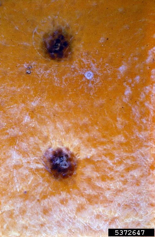
  • Citrus Black Spot Wikipedia
    Recommended Chemical Controls for Citrus Black Spot [28] Pesticide FRAC MOA2 Mature Trees Rate/Acre1 copper fungicide M1 use label rate Abound 2.08F3 11 12.4-15.4 fl oz.
  • ←
  • 1
  • 2
  • ...
  • 157
  • 158
  • 159
  • 160
  • 161
  • 162
  • 163
  • ...
  • 188
  • 189
  • →

FindZebra

contact@findzebra.com