Binswanger's disease is a type of dementia caused by widespread, microscopic areas of damage to the deep layers of white matter in the brain. Most affected people experience progressive memory loss and deterioration of intellectual abilities (dementia); urinary urgency or incontinence; and an abnormally slow, unsteady gait (style of walking). While there is no cure, the progression of Binswanger's disease can be slowed with healthy lifestyle choices. Treatment is based on the signs and symptoms present in each person.
Joubert syndrome (JS) and related disorders (JSRD) are a group of developmental delay/multiple congenital anomaly syndromes in which the mandatory feature is the ``molar tooth sign'' (MTS), a complex midbrain-hindbrain malformation recognizable on brain imaging. ... Differential diagnosis Differential diagnosis must consider in particular the other ciliopathies (such as nephronophthisis, Senior-Loken syndrome, and Bardet-Biedl syndrome; see these terms), distinct cerebellar and brainstem congenital defects and disorders with cerebro-oculo-renal manifestations.
Acorea, microphthalmia and cataract syndrome Specialty Ophthalmology Acorea, microphthalmia and cataract syndrome is a rare genetically inherited condition. [1] Contents 1 Presentation 2 Genetics 3 Diagnosis 4 Treatment 5 References Presentation [ edit ] Acorea or fibrous occlusion of the pupil, microphthalmia and cataracts are present in both eyes. ... You can help by adding to it . ( December 2017 ) References [ edit ] ^ Kondo H, Tahira T, Yamamoto K, Tawara A (2013) Familial acorea, microphthalmia and cataract syndrome. Br J Ophthalmol v t e Congenital malformations and deformations of eyes Adnexa Eyelid Ptosis Ectropion Entropion Distichia Blepharophimosis Ablepharon Marcus Gunn phenomenon Lacrimal apparatus Congenital lacrimal duct obstruction Globe Entire eye Anophthalmia ( Cystic eyeball , Cryptophthalmos ) Microphthalmia Lens Ectopia lentis Aphakia Iris Aniridia Anterior segment Axenfeld–Rieger syndrome Cornea Keratoglobus Megalocornea Other Buphthalmos Coloboma ( Coloboma of optic nerve ) Hydrophthalmos Norrie disease v t e Medicine Specialties and subspecialties Surgery Cardiac surgery Cardiothoracic surgery Colorectal surgery Eye surgery General surgery Neurosurgery Oral and maxillofacial surgery Orthopedic surgery Hand surgery Otolaryngology ENT Pediatric surgery Plastic surgery Reproductive surgery Surgical oncology Transplant surgery Trauma surgery Urology Andrology Vascular surgery Internal medicine Allergy / Immunology Angiology Cardiology Endocrinology Gastroenterology Hepatology Geriatrics Hematology Hospital medicine Infectious disease Nephrology Oncology Pulmonology Rheumatology Obstetrics and gynaecology Gynaecology Gynecologic oncology Maternal–fetal medicine Obstetrics Reproductive endocrinology and infertility Urogynecology Diagnostic Radiology Interventional radiology Nuclear medicine Pathology Anatomical Clinical pathology Clinical chemistry Cytopathology Medical microbiology Transfusion medicine Other Addiction medicine Adolescent medicine Anesthesiology Dermatology Disaster medicine Diving medicine Emergency medicine Mass gathering medicine Family medicine General practice Hospital medicine Intensive care medicine Medical genetics Narcology Neurology Clinical neurophysiology Occupational medicine Ophthalmology Oral medicine Pain management Palliative care Pediatrics Neonatology Physical medicine and rehabilitation PM&R Preventive medicine Psychiatry Addiction psychiatry Radiation oncology Reproductive medicine Sexual medicine Sleep medicine Sports medicine Transplantation medicine Tropical medicine Travel medicine Venereology Medical education Medical school Bachelor of Medicine, Bachelor of Surgery Bachelor of Medical Sciences Master of Medicine Master of Surgery Doctor of Medicine Doctor of Osteopathic Medicine MD–PhD Related topics Alternative medicine Allied health Dentistry Podiatry Pharmacy Physiotherapy Molecular oncology Nanomedicine Personalized medicine Public health Rural health Therapy Traditional medicine Veterinary medicine Physician Chief physician History of medicine Book Category Commons Wikiproject Portal Outline
Achondrogenesis is a group of severe disorders that are present from birth and affect the development of cartilage and bone. Infants with achondrogenesis usually have a small body, extremely short arms and legs, other skeletal abnormalities, and underdeveloped lungs. There are at least three forms of achondrogenesis, type 1A, type 1B and type 2. Achondrogenesis is usually diagnosed during pregnancy by ultrasound and genetic testing is used to distinguish between the three types. Type 1A and 1B achondrogenesis are both inherited in an autosomal recessive pattern.
A number sign (#) is used with this entry because of evidence that achondrogenesis type IA (ACG1A) is caused by homozygous or compound heterozygous mutation in the TRIP11 gene (604505) on chromosome 14q32. Description The term achondrogenesis has been used to characterize the most severe forms of chondrodysplasia in humans, invariably lethal before or shortly after birth. Achondrogenesis type I is a severe chondrodystrophy characterized radiographically by deficient ossification in the lumbar vertebrae and absent ossification in the sacral, pubic and ischial bones and clinically by stillbirth or early death (Maroteaux and Lamy, 1968; Langer et al., 1969). In addition to severe micromelia, there is a disproportionately large cranium due to marked edema of soft tissues. Classification of Achondrogenesis Achondrogenesis was traditionally divided into 2 types: type I (Parenti-Fraccaro) and type II (Langer-Saldino).
A rare group of lethal skeletal dysplasias characterized by an endochondral ossification deficiency that leads to dwarfism with extreme micromelia, a small thorax, a prominent abdomen, anasarca and polyhydramnios. There are three types of achondrogenesis that exist and that differ clinically, radiologically, histologically and genetically: achondrogensis type 1a, type 1b and type 2.
A number sign (#) is used with this entry because of evidence that brachydactyly-syndactyly syndrome is caused by heterozygous mutation in the HOXD13 gene (142989) on chromosome 2q31. ... Clinical Features Zhao et al. (2007) described a Han Chinese family in which 23 affected individuals in 6 generations exhibited a complex brachydactyly-syndactyly syndrome. Digital photographs and radiographs were taken for 16 and 13 of them, respectively. ... Brachydactyly/Syndactyly/Oligodactyly Syndrome Ibrahim et al. (2013) studied a girl with a complex brachydactyly-syndactyly-oligodactyly phenotype, who was born with normal measurements and had normal psychomotor development. ... Molecular Genetics In a large Han Chinese family segregating brachydactyly-syndactyly syndrome, Zhao et al. (2007) found deletion of 21 basepairs in the HOXD13 gene (142989.0010) in affected members.
Brachydactyly-syndactyly, Zhao type is a recently described syndrome associating a brachydactyly type A4 (short middle phalanges of the 2nd and 5th fingers and absence of middle phalanges of the 2nd to 5th toes) and a syndactyly of the 2nd and 3rd toes. Metacarpals and metatarsals anomalies are common. Epidemiology This syndrome has been described in two families.
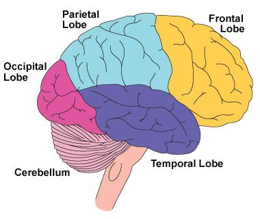
The fact that many of the dysexecutive syndrome symptoms can occur alone has led some researchers [8] to suggest that the symptoms should not be labelled as a "syndrome" as such. ... Frontal disinhibition syndrome, Rett syndrome and attention deficit hyperactivity disorder [13] It is produced from frontal lobe damage often due to tumors. ... Retrieved 2008-07-02 . ^ a b "Frontal Lobe Syndrome. FLS information. Frontal Lobe Lesions | Patient" . ... Retrieved 2016-01-30 . ^ "Foster Kennedy's Syndrome. FKS information. Patient | Patient" . ... "Frontal lobe disinhibition, Rett syndrome and attention deficit hyperactivity disorder".
The phenotypic spectrum of MED12 -related disorders, which is still being defined, includes at a minimum the phenotypes of FG syndrome type 1 (FGS1), Lujan syndrome (LS), and X-linked Ohdo syndrome. ... Diagnosis Suggestive Findings An MED12 -related disorder should be suspected in an individual with a phenotype associated with FG syndrome type 1, Lujan syndrome, or X-linked Ohdo syndrome. ... FG syndrome type 1 is also referred to as Opitz-Kaveggia syndrome [Risheg et al 2007]. ... Inheritance is X-linked. Phelan-McDermid syndrome . (22q13.3 deletion syndrome) Common features seen in both FGS1 and 22q13.3 deletion syndrome include hypotonia, intellectual disability, and delayed speech. ... MID1 is the only gene associated with X-linked Opitz G/BBB syndrome. Rubinstein-Taybi syndrome (RSTS).
Lethal white syndrome Other names overo lethal white syndrome (OLWS), lethal white overo (LWO), overo lethal white foal syndrome (OLWFS) Healthy horse exhibiting the frame overo pattern. ... Prevention Avoid breeding heterozygous frame horses to each other Treatment None Lethal white syndrome ( LWS ), also called overo lethal white syndrome ( OLWS ), lethal white overo ( LWO ), and overo lethal white foal syndrome ( OLWFS ), is an autosomal genetic disorder most prevalent in the American Paint Horse . ... "The Impact of the Mutation Causing Overo Lethal White Syndrome on White Patterning in Horses" (PDF) . ... "Endothelin receptor B polymorphism associated with lethal white foal syndrome in horses" . Mammalian Genome . ... Santschi (1998-07-01). "Stalking the Lethal White Syndrome" . Paint Horse Journal . American Paint Horse Association.
Meralgia paresthetica Other names Bernhardt - Roth syndrome [1] Innervation of lateral cutaneous nerve of the thigh (shaded area) on the right leg. ... The disorder has also been nicknamed skinny pants syndrome , [3] in reference to a rise in teenagers wearing skin-tight trousers. ... "Meralgia paraesthetica (Bernhardt-Roth syndrome)" . Journal of Neurology, Neurosurgery & Psychiatry . 77 (1): 84. doi : 10.1136/jnnp.2005.072363 . ... PMID 16361600 . ^ IASP, XXXI: LOCAL SYNDROMES IN THE LEG OR FOOT: PAIN OF NEUROLOGICAL ORIGIN Archived 2012-12-19 at the Wayback Machine , 2012 ^ "Make your pencil jeans less dangerous to your health" . ... Retrieved 2007-04-09 . ^ a b c Meralgia Paresthetica orthoped/416 at eMedicine ^ a b Meralgia Paresthetica neuro/590 at eMedicine External links [ edit ] Classification D ICD - 10 : G57.1 ICD - 9-CM : 355.1 MeSH : C537458 C537458, C537458 DiseasesDB : 31968 External resources eMedicine : neuro/590 orthoped/416 , pmr/76 Patient UK : Meralgia paraesthetica Meralgia Paresthetica at eMedicine.com v t e Diseases relating to the peripheral nervous system Mononeuropathy Arm median nerve Carpal tunnel syndrome Ape hand deformity ulnar nerve Ulnar nerve entrapment Froment's sign Ulnar tunnel syndrome Ulnar claw radial nerve Radial neuropathy Wrist drop Cheiralgia paresthetica long thoracic nerve Winged scapula Backpack palsy Leg lateral cutaneous nerve of thigh Meralgia paraesthetica tibial nerve Tarsal tunnel syndrome plantar nerve Morton's neuroma superior gluteal nerve Trendelenburg's sign sciatic nerve Piriformis syndrome Cranial nerves See Template:Cranial nerve disease Polyneuropathy and Polyradiculoneuropathy HMSN Charcot–Marie–Tooth disease Dejerine–Sottas disease Refsum's disease Hereditary spastic paraplegia Hereditary neuropathy with liability to pressure palsy Familial amyloid neuropathy Autoimmune and demyelinating disease Guillain–Barré syndrome Chronic inflammatory demyelinating polyneuropathy Radiculopathy and plexopathy Brachial plexus injury Thoracic outlet syndrome Phantom limb Other Alcoholic polyneuropathy Other General Complex regional pain syndrome Mononeuritis multiplex Peripheral neuropathy Neuralgia Nerve compression syndrome
Meralgia paresthetica is a condition characterized by numbness, tingling, and a burning pain in the outer thigh. Symptoms may worsen after walking or standing. The condition usually affects only one side of the body, but both sides may be affected in up to 20% of cases. Meralgia paresthetica is caused by compression of the lateral femoral cutaneous nerve, a sensory nerve to the skin on the outer thigh. Compression may be associated with various causes such as wearing tight clothing or a heavy tool belt, diabetes, nerve injury during local or regional surgery, weight gain, pregnancy, seatbelt injury, or rarely, a mass pressing on the nerve. Treatment is based on the symptoms and severity in each person. Treatment for mild symptoms may include removing the cause of nerve compression, which may involve weight loss or wearing loose clothing.
Overview Meralgia paresthetica (also known as lateral femoral cutaneous nerve entrapment) is a condition characterized by tingling, numbness and burning pain in your outer thigh. It's caused by compression of the nerve that provides sensation to the skin covering your thigh. Tight clothing, obesity or weight gain, and pregnancy are common causes of meralgia paresthetica. However, meralgia paresthetica can also be due to local trauma or a disease, such as diabetes. In most cases, you can relieve meralgia paresthetica with conservative measures, such as wearing looser clothing.
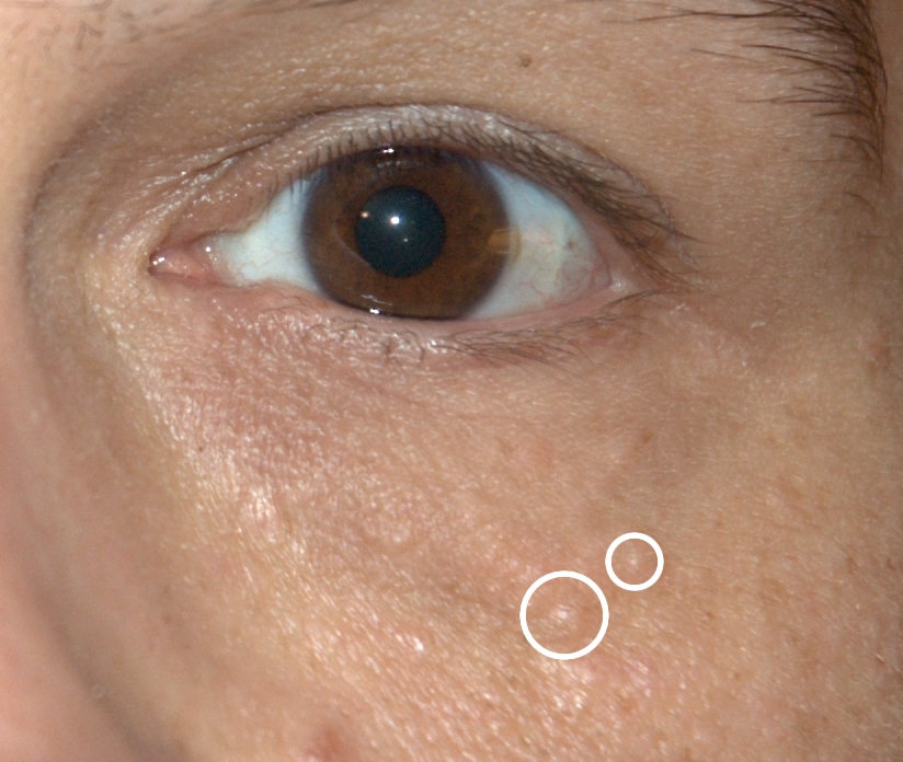
Presentation [ edit ] Associated syndromes [ edit ] Syringomas can be found in association with other symptoms as part of a syndrome . Hailey–Hailey disease (also known as familial benign chronic pemphigus) is a blistering disease that can also include syringomas. [5] Several systemic syndromes have also been associated with syringoma including diabetes mellitus , Down syndrome , Brooke-Spiegler and Nicolau-Balus . ... The incidence of syringomas has been reported in up to 40 percent of people with Down syndrome and can be associated with a condition calcinosis cutis which requires prompt medical attention. Brooke-Spiegler syndrome is a rare autosomal dominant syndrome with cutaneous manifestations including syringomas and trichoepitheliomas . Nicolau-Balus syndrome is a rare autosomal dominant disorder consisting of atrophoderma vermiculata and syringomas.
Progressive enlargement of the globe or "buphthalmos" usually does not occur after age three to four years [Ho & Walton 2004, Allingham et al 2005]. Conditions/syndromes associated with infantile glaucoma. A number of well-recognized conditions and syndromes may present with infantile glaucoma, along with other ocular and/or systemic findings. Some conditions may not be compatible with life (e.g., trisomy 13, trisomy 18, Walker-Warburg syndrome, and Zellweger Syndrome); others may be less severe or confined only to the eye. It is important to establish the diagnosis of an associated syndrome because of the implications for genetic counseling and treatment (see Table 2). Table 2. Conditions/Syndromes Associated with Infantile Glaucoma View in own window Disorder Gene(s) MOI Clinical Features Eye Findings Other Aniridia PAX6 WT1 1 AD Complete or partial iris hypoplasia w/associated foveal hypoplasia, resulting in reduced visual acuity & nystagmus Presents in early infancy Frequently associated w/other ocular abnormalities, often of later onset, incl cataract, glaucoma, & corneal opacification& vascularization May occur either as an isolated ocular abnormality w/out systemic involvement or as part of WAGR syndrome 1 Anterior segment dysgenesis syndromes (e.g., Peters Plus syndrome) See footnote 2 Phenotypically & genotypically distinct from PCG in general, but severe or advanced PCG can be difficult to distinguish clinically from some of the anterior segment dysgenesis syndromes; e.g., Peters anomaly Peters Plus syndrome: developmental delay, mild to severe ID, cleft lip, cleft palate Axenfeld-Rieger anomaly (anterior segment disorder) FOXC1 PITX2 AD Presents w/posterior embryotoxon & (variably) iris strands adherent to Schwalbe's line, iris hypoplasia, focal iris atrophy, & ectropion uveae.
Severe combined immunodeficiency (SCID) due to adenosine deaminase (ADA) deficiency is a form of SCID characterized by profound lymphopenia and very low immunoglobulin levels of all isotypes resulting in severe and recurrent opportunistic infections. Epidemiology SCID due to ADA deficiency accounts for 10-15% of all cases of SCID. Its annual incidence is estimated to be between 1/200,000 and 1/1,000,000 live births. Both males and females are affected. Clinical description SCID due to ADA deficiency has a variable clinical presentation. The most common form presents in infancy with severe and recurrent opportunistic infections (including respiratory tract infections and candidiasis), failure to thrive, and usually results in early death.
Rauch et al. (1999) reported 2 sisters with a syndrome of severe developmental delay, ataxia, impaired social interaction, seizure disorder with early onset but without epileptiform electroencephalographic changes, and a striking light-fixating behavior which was associated with retinal cone dystrophy. ... There was no evidence of regression in the patients of Rauch et al. (1999) and hyperreflexia seen in the sisters is absent in the EFMR syndrome. All patients with EFMR remained ambulatory.