A rare primary bone dysplasia with decreased bone density disorder characterized by multiple doughnut-shaped hyperostotic or osteosclerotic clavarial lesions (manifesting with cranial lumps) associated with numerous pathologic fractures, elevated serum alkaline phosphatase levels and osteopenia.
A number sign (#) is used with this entry because of evidence that calvarial doughnut lesions with bone fragility (CDL) and CDL with spondylometaphyseal dysplasia (CDLSMD) are caused by heterozygous mutation in the SGMS2 gene (611574) on chromosome 4q25. Description Calvarial doughnut lesions with bone fragility (CDL) is characterized by low bone mineral density, multiple spinal and peripheral fractures beginning in childhood, and sclerotic doughnut-shaped lesions in the cranial bones. Some more severely affected individuals exhibit neonatal onset of fractures, severe short stature, marked cranial sclerosis, and spondylometaphyseal dysplasia (CDLSMD) (Pekkinen et al., 2019). Clinical Features Bartlett and Kishore (1976) described multiple hyperostotic or osteosclerotic lesions of the calvaria in a man and 3 of his sons, each by a different mother. One of the affected sons also experienced recurrent facial nerve palsy.
Adrenocortical adenomas are classified as ACTH -independent disorders, and are commonly associated with conditions linked to hyperadrenalism such as Cushing's syndrome ( hypercortisolism ) or Conn's syndrome ( hyperaldosteronism ), which is also known as primary aldosteronism . [1] In addition, recent case reports further support the affiliation of adrenocortical adenomas with hyperandrogenism or florid hyperandrogenism which can cause hyperandrogenic hirsutism in females. [2] " Cushing's syndrome " differs from the " Cushing's disease " even though both conditions are induced by hypercortisolism . The term " Cushing's disease " refers specifically to "secondary hypercortisolism" classified as " ACTH-dependent Cushing's syndrome" caused by pituitary adenomas . In contrast, "Cushing's syndrome" refers specifically to "primary hypercortisolism" classified as " ACTH-independent Cushing's syndrome" caused by adrenal adenomas . ... Prevalence: Female > Male More common in adults Relatively earlier onset in females (ages ≤ 20) than males (ages ≤ 30) Most common cause of ACTH -independent Cushing's syndrome See also [ edit ] Hyperplasia Adrenal tumor Cushing's syndrome Conn's syndrome Hypercortisolism Hyperaldosteronism Hyperandrogenism Adrenal gland Adrenal paraganglioma Adrenal Pheochromocytoma Adrenal ganglioneuroma References [ edit ] ^ "Definition: adrenocortical adenoma from Online Medical Dictionary" . ^ LaVoie, Melanie; Constantinides, Vasilis; Robin, Noel; Kyriacou, Angelos (30 July 2018). ... "Bilateral Adrenocortical Adenomas along with Virilization and Cushing's Syndrome" . Internal Medicine . 58 (3): 405–409. doi : 10.2169/internalmedicine.0790-18 .
High levels of these hormones can lead to complications, including primary aldosteronism , Cushing's syndrome and other medical conditions. Functioning adrenal adenomas may be treated with surgery and/or medications.
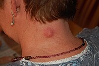
Overview Epidermoid (ep-ih-DUR-moid) cysts are noncancerous small bumps beneath the skin. They can appear anywhere on the skin, but are most common on the face, neck and trunk. Epidermoid cysts are slow growing and often painless, so they rarely cause problems or need treatment. You might choose to have a cyst removed by a doctor if its appearance bothers you or if it's painful, ruptured or infected. Many people refer to epidermoid cysts as sebaceous cysts, but they're different.
A number sign (#) is used with this entry because of evidence that congenital interstitial lung disease with nephrotic syndrome and epidermolysis bullosa (ILNEB) is caused by homozygous mutation in the ITGA3 gene (605025) on chromosome 17q21. ... Patients exhibit a multiorgan disorder that includes congenital interstitial lung disease, nephrotic syndrome, and epidermolysis bullosa. The respiratory and renal features predominate, and lung involvement accounts for the lethal course of the disease (summary by Has et al., 2012). Clinical Features Has et al. (2012) described 3 unrelated children, a boy and 2 girls, who had congenital interstitial lung disease, nephrotic syndrome, and mild epidermolysis bullosa. ... At 2 weeks of age, he developed renal failure and nephrotic syndrome, and peritoneal dialysis was initiated. ... Molecular Genetics In an Italian boy with congenital interstitial lung disease, nephrotic syndrome, and mild epidermolysis bullosa, who died at 7.5 months of age of a lung infection, Has et al. (2012) excluded mutation in the NPHS2 (604766) and WT1 (607102) genes as a cause of the nephrotic syndrome and also ruled out mutation in the CFTR (602421) and ABCA3 (601615) genes as a cause of the interstitial lung disease.
Congenital nephrotic syndrome-interstitial lung disease-epidermolysis bullosa syndrome is a life-threatening multiorgan disorder which develops in the first months of life, presenting with respiratory distress and proteinuria in the nephrotic range, and leading to severe interstitial lung disease and renal failure.
A clinically isolated syndrome ( CIS ) is a clinical situation of an individual's first neurological episode, caused by inflammation or demyelination of nerve tissue. ... Brain lesions associated with a clinically isolated syndrome may be indicative of several neurological diseases, like multiple sclerosis (MS) or neuromyelitis optica . ... References [ edit ] ^ "Clinically Isolated Syndrome (CIS)" . Library . Archived from the original on 2007-12-06 . ... External links [ edit ] Diagnosing MS in Clinically Isolated Syndrome—The Role of MRI v t e Multiple sclerosis and other demyelinating diseases of the central nervous system Signs and symptoms Ataxia Depression Diplopia Dysarthria Dysphagia Fatigue Incontinence Nystagmus Optic neuritis Pain Uhthoff's phenomenon Investigations and diagnosis Multiple sclerosis diagnosis McDonald criteria Poser criteria Clinical Clinically isolated syndrome Expanded Disability Status Scale Serological and CSF Oligoclonal bands Radiological Radiologically isolated syndrome Lesional demyelinations of the central nervous system Dawson's fingers Approved [ by whom? ] treatment Management of multiple sclerosis Alemtuzumab Cladribine Dimethyl fumarate Fingolimod Glatiramer acetate Interferon beta-1a Interferon beta-1b Mitoxantrone Natalizumab Ocrelizumab Ozanimod Siponimod Teriflunomide Other treatments Former Daclizumab Multiple sclerosis research Demyleinating diseases Autoimmune Multiple sclerosis Neuromyelitis optica Diffuse myelinoclastic sclerosis Inflammatory Acute disseminated encephalomyelitis MOG antibody disease Balo concentric sclerosis Marburg acute multiple sclerosis Neuromyelitis optica Diffuse myelinoclastic sclerosis Tumefactive multiple sclerosis Experimental autoimmune encephalomyelitis Hereditary Adrenoleukodystrophy Alexander disease Canavan disease Krabbe disease Metachromatic leukodystrophy Pelizaeus–Merzbacher disease Leukoencephalopathy with vanishing white matter Megalencephalic leukoencephalopathy with subcortical cysts CAMFAK syndrome Other Central pontine myelinolysis Marchiafava–Bignami disease Mitochondrial DNA depletion syndrome Other List of multiple sclerosis organizations List of people with multiple sclerosis Multiple sclerosis drug pipeline Pathophysiology
Prion disease characterized by adult onset of memory loss, dementia, ataxia, and pathologic deposition of amyloid-like plaques in the brain Not to be confused with Gerstmann syndrome . Gerstmann–Sträussler–Scheinker syndrome A person with inherited prion disease has cerebellar atrophy. ... Causes prion Prognosis (AVERAGE) 5-6 years from diagnosis Gerstmann–Sträussler–Scheinker syndrome ( GSS ) is an extremely rare, usually familial, fatal neurodegenerative disease that affects patients from 20 to 60 years in age. ... "Gerstmann–Sträussler–Scheinker syndrome, fatal familial insomnia, and kuru: a review of these less common human transmissible spongiform encephalopathies". ... Retrieved April 6, 2011 . ^ Tesar A, Matej R, Kukal J, Johanidesova S, Rektorova I, Vyhnalek M, Keller J, Eliasova I, Parobkova E, Smetakova M, Musova Z, Rusina R (2019) Clinical variability in P102L Gerstmann-Sträussler-Scheinker Syndrome. Ann Neurol ^ Arata H, Takashima H, Hirano R, et al. ... "Early clinical signs and imaging findings in Gerstmann–Sträussler–Scheinker syndrome (Pro102Leu)". Neurology . 66 (11): 1672–8. doi : 10.1212/01.wnl.0000218211.85675.18 .
Contents 1 Signs and symptoms 1.1 Clinical features 1.2 Associated conditions 2 Cause 3 Mechanism 4 Diagnosis 4.1 Histology 5 Management 6 See also 7 References 8 External links Signs and symptoms [ edit ] Clinical features [ edit ] NME features a characteristic skin eruption of red patches with irregular borders, intact and ruptured vesicles , and crust formation. [1] It commonly affects the limbs and skin surrounding the lips, although less commonly the abdomen, perineum , thighs, buttocks, and groin may be affected. [1] Frequently these areas may be left dry or fissured as a result. [1] All stages of lesion development may be observed synchronously. [2] The initial eruption may be exacerbated by pressure or trauma to the affected areas. [1] Associated conditions [ edit ] William Becker first described an association between NME and glucagonoma in 1942 [2] [3] and since then, NME has been described in as many as 70% of persons with a glucagonoma. [4] NME is considered part of the glucagonoma syndrome , [5] which is associated with hyperglucagonemia , diabetes mellitus , and hypoaminoacidemia . [2] When NME is identified in the absence of a glucagonoma, it may be considered "pseudoglucagonoma syndrome". [6] Less common than NME with glucagonoma, pseudoglucagonoma syndrome may occur in a number of systemic disorders: [7] Celiac disease Ulcerative colitis Crohn's disease Hepatic cirrhosis Hepatocellular carcinoma Lung cancer , including small cell lung cancer Tumors that secrete insulin - or insulin-like growth factor 2 Duodenal cancer Cause [ edit ] The cause of NME is unknown, although various mechanisms have been suggested. ... Archives of Dermatology and Syphilology . 45 (6): 1069–1080. doi : 10.1001/archderm.1942.01500120037004 . ^ van Beek AP, de Haas ER, van Vloten WA, Lips CJ, Roijers JF, Canninga-van Dijk MR (November 2004). "The glucagonoma syndrome and necrolytic migratory erythema: a clinical review" . ... "Histologic variation in the skin lesions of the glucagonoma syndrome". The American Journal of Surgical Pathology . 10 (7): 445–53. doi : 10.1097/00000478-198607000-00001 . ... External links [ edit ] Classification D MeSH : D058568 DiseasesDB : 8833 SNOMED CT : 15576007 External resources eMedicine : derm/168 v t e Urticaria and erythema Urticaria ( acute / chronic ) Allergic urticaria Urticarial allergic eruption Physical urticaria Cold urticaria Familial Primary cold contact urticaria Secondary cold contact urticaria Reflex cold urticaria Heat urticaria Localized heat contact urticaria Solar urticaria Dermatographic urticaria Vibratory angioedema Pressure urticaria Cholinergic urticaria Aquagenic urticaria Other urticaria Acquired C1 esterase inhibitor deficiency Adrenergic urticaria Exercise urticaria Galvanic urticaria Schnitzler syndrome Urticaria-like follicular mucinosis Angioedema Episodic angioedema with eosinophilia Hereditary angioedema Erythema Erythema multiforme / drug eruption Erythema multiforme minor Erythema multiforme major Stevens–Johnson syndrome , Toxic epidermal necrolysis panniculitis ( Erythema nodosum ) Acute generalized exanthematous pustulosis Figurate erythema Erythema annulare centrifugum Erythema marginatum Erythema migrans Erythema gyratum repens Other erythema Necrolytic migratory erythema Erythema toxicum Erythroderma Palmar erythema Generalized erythema v t e Paraneoplastic syndromes Endocrine Hypercalcaemia SIADH Zollinger–Ellison syndrome Cushing's syndrome Hematological Multicentric reticulohistiocytosis Nonbacterial thrombotic endocarditis Neurological Paraneoplastic cerebellar degeneration Encephalomyelitis Limbic encephalitis Opsoclonus Polymyositis Transverse myelitis Lambert–Eaton myasthenic syndrome Anti-NMDA receptor encephalitis Musculoskeletal Dermatomyositis Hypertrophic osteopathy Mucocutaneous reactive erythema Erythema gyratum repens Necrolytic migratory erythema papulosquamous Acanthosis nigricans Ichthyosis acquisita Acrokeratosis paraneoplastica of Bazex Extramammary Paget's disease Florid cutaneous papillomatosis Leser-Trélat sign Pityriasis rotunda Tripe palms Other Febrile neutrophilic dermatosis Pyoderma gangrenosum Paraneoplastic pemphigus
A rare genetic disease characterized by choanal atresia and early onset of lymphedema of the lower extremities. Additional reported features include facial dysmorphism (hypertelorism, broad forehead, smooth philtrum, unilateral low-set ear, and high-arched palate), hypoplastic nipples, and pectus excavatum.
A number sign (#) is used with this entry because of evidence that choanal atresia and lymphedema (CATLPH) is caused by homozygous mutation in the PTPN14 gene (603155) on chromosome 1q32-q41. Clinical Features Qazi et al. (1982) reported a consanguineous Yemenite family in which a brother and sister and their paternal aunt had posterior choanal atresia. The boy also had a high-arched palate, hypoplastic nipples, and mild pectus excavatum, and the aunt had a high-arched palate. All 4 parents of the 3 affected persons could be traced to a common ancestral couple 2 or 3 generations earlier. Har-El et al. (1991) provided follow-up of the family reported by Qazi et al. (1982).
Progressive sensorineural hearing loss - hypertrophic cardiomyopathy is an extremely rare disorder described in one family to date that is characterized by progressive, late onset, autosomal dominant sensorineural hearing loss, QT interval prolongation, and mild cardiac hypertrophy.
A number sign (#) is used with this entry because of evidence that autosomal dominant nonsyndromic deafness-22 (DFNA22) is caused by heterozygous mutation in the myosin VI gene (MYO6; 600970) on chromosome 6q14. Clinical Features Melchionda et al. (2001) studied a large Italian kindred in which many members were affected with autosomal dominant nonsyndromic sensorineural hearing loss. Hearing loss was progressive and postlingual, with onset during childhood (8 to 10 years of age at onset of symptoms; 6 to 8 years of age at onset of first audiometric abnormalities). By the age of approximately 50 years, affected individuals invariably had profound sensorineural deafness. Vestibular and/or visual involvement was excluded in all affected individuals.
Cutaneous photosensitivity and lethal colitisis is a rare inflammatory bowel disease (see this term) characterized by early cutaneous photosensitivity manifesting by sun-induced facial erythematous and vesicular lesions and severe recurent colitis which lead to untreatable diarrhea. There have been no further descriptions in the literature since 1991.
Labrune et al. (1991) described early cutaneous photosensitivity and severe colitis in 3 of 4 sibs born to unrelated parents. All 3 affected sibs died from refractory diarrhea. The fourth sib, who had a different father, was normal. The first infant developed erythematous and vesicular lesions of the face after a 6-hour exposure to sun at the age of 2 weeks and died at the age of 6 months. The second sib, a girl, died at the age of 20 months after a similar course. Porphyria was excluded. Normal plasma zinc concentration in 1 of the sibs strongly argued against the diagnosis of acrodermatitis enteropathica (201100).
A rare hematologic disease characterized by high serum viscosity due to polyclonal expansion of immunoglobulins, most commonly in the context of Waldenström's macroglobulinemia, as well as a variety of disorders of immune dysregulation. Patients present with signs and symptoms involving multiple organs, such as bleeding diathesis, mucosal bleeding, retinal hemorrhage, headache, stroke, pulmonary hypertension, and congestive heart failure.
A rare genetic disease characterized by congenital cataract, neonatal hepatic failure and cholestatic jaundice, and global developmental delay. Neonatal death due to progressive liver failure has been reported.
A complex form of hereditary spastic paraplegia characterized by spastic paraplegia, demyelinating peripheral sensorimotor neuropathy, poikiloderma (manifesting with loss of eyebrows and eyelashes in childhood in addition to delicate, smooth, and wasted skin) and distal amyotrophy (presenting after puberty). There have been no further descriptions in the literature since 1992.
In a family of Portuguese extraction, Antinolo et al. (1992) described 7 individuals in 4 sibships in 3 generations with a disorder comprising spastic paraplegia, demyelinating peripheral neuropathy, and poikiloderma, manifested by 'delicate, smooth, and wasted' skin from an early age and loss of eyebrows and eyelashes at age 3 years. Electroneurophysiologic studies demonstrated motor and sensory neuropathy. Sural nerve biopsy showed onion bulb formations, indicating demyelination of peripheral nerves. Distal amyotrophy was evident after puberty. There were 2 affected males but no male-to-male transmission. Neuro - Spastic paraplegia - Demyelinating peripheral neuropathy Inheritance - Autosomal dominant Lab - Motor and sensory neuropathy on electroneurophysiologic studies - Onion bulb formations on sural nerve biopsy Hair - Loss of eyebrows and eyelashes by age 3 years Skin - Poikiloderma - Delicate, smooth, wasted skin Muscle - Distal amyotrophy ▲ Close
An extremely rare genetic congenital heart disease characterized by the presence of atrial septal defect, mostly of the ostium secundum type, associated with conduction anomalies like atrioventricular block, atrial fibrillation or right bundle branch block.
The family of Weil and Allenstein (1961) probably represented an example of this syndrome. The occurrence of other forms of congenital heart disease in this syndrome was suggested by the family reported by Pease et al. (1976). ... The patient later developed sick sinus syndrome and required permanent pacemaker implantation. His mother had the same mutation and ASD with atrial fibrillation; her elder sister had ASD secundum with AV block and sick sinus syndrome requiring permanent pacemaker implantation, and a nephew had ASD with AV block.
A rare systemic disease characterized by febrile illness (body temperature >38.3°C on several occasions) or inflammation (elevated serum C-reactive protein and erythrocyte sedimentation rate) lasting at least three weeks and for which no specific diagnosis is achieved despite extended diagnostics.
A rare genetic bone disease characterized by short stature, bilateral congenital hip dislocation, radial head dislocation, carpal coalition, scoliosis, pes cavus, and atlantoaxial subluxation. Dysmorphic facial features include broad forehead, broad nasal bridge, hypertelorism, and mild midface hypoplasia. Association with bilateral sensorineural hearing loss has also been described.
A number sign (#) is used with this entry because of evidence that Steel syndrome (STLS) is caused by homozygous or compound heterozygous mutation in the COL27A1 gene (608461) on chromosome 9q32. Description Steel syndrome is characterized by characteristic facies, dislocated hips and radial heads, carpal coalition (fusion of carpal bones), short stature, scoliosis, and cervical spine anomalies. ... There was no evidence that any of these 5 individuals had received a clinical diagnosis of Steel syndrome. Among the 34 heterozygous individuals below the age of 55 years (mean 41.8 years), none had short stature, and none had hip or radial head dislocations. ... By exome sequencing in a 5-year-old girl, born to nonconsanguineous Indian parents, with Steel syndrome, Kotabagi et al. (2017) identified compound heterozygosity for a nonsense (R707X; 608461.0002) and a frameshift (608461.0003) mutation in the COL27A1 gene. ... By exome sequencing in a 3-year-old girl with Steel syndrome, who was born to first-cousin parents from the United Arab Emirates, Gariballa et al. (2017) identified a homozygous splice site mutation (608461.0004) in the COL27A1 gene.
A rare sterol biosynthesis disorder characterized by microcephaly, bilateral congenital cataract, mild developmental delay, growth delay with short stature, psoriasiform dermatitis of variable severity, and immune dysregulation. Behavioral disorder, joint contractures, and arthralgia have also been described.
A number sign (#) is used with this entry because of evidence that microcephaly, congenital cataract, and psoriasiform dermatitis (MCCPD) is caused by homozygous or compound heterozygous mutation in the SC4MOL gene (MSMO1; 607545) on chromosome 4q32. Description SC4MOL deficiency represents an inborn error of cholesterol metabolism that is characterized by accumulation of a large amount of methylsterols, particularly dimethylsterols, in affected patients. The associated features of immune dysregulation, skin disease, and growth delay can be at least partially corrected with cholesterol and statin supplements (He et al., 2014). Clinical Features He et al. (2011) reported a 15-year-old girl who presented at age 13 years for evaluation of severe ichthyosiform erythroderma affecting the entire body but sparing the palms. Her skin was normal at birth, but periumbilical dermatitis was noted when she was 2 years old; dermatitis then spread to her back and trunk and ultimately to the remainder of her body by age 6.