-
Feingold Syndrome 1
Gene_reviews
Approximately 60% of individuals with Feingold syndrome 1 have an affected parent; the proportion of FS1 caused by a de novo MYCN pathogenic variant is unknown. ... Diagnosis Suggestive Findings Feingold syndrome 1 (FS1) should be suspected in individuals with the following clinical findings [Marcelis et al 2008]. ... Nomenclature Terms used in the past for Feingold syndrome 1: Microcephaly-oculo-digito-esophageal-duodenal syndrome Microcephaly mesobrachyphalangy tracheoesophageal fistula syndrome Microcephaly-digital anomalies-normal intelligence syndrome Prevalence Prevalence is unknown; FS1 is likely rare. ... Feingold syndrome 2 is caused by hemizygous deletions of chromosome 13q31.3 including MIR17HG [Tassano et al 2013]. 2. To date, seven individuals with Feingold syndrome 2 have been described [de Pontual et al 2011, Ganjavi et al 2014, Muriello et al 2019]. 3.
-
Menstrual Disorder
Wikipedia
Retrieved 2020-07-31 . ^ Dickerson LM, Mazyck PJ, Hunter MH (April 2003). "Premenstrual syndrome". American Family Physician . 67 (8): 1743–52. PMID 12725453 . ^ Yonkers KA, O'Brien PM, Eriksson E (April 2008). "Premenstrual syndrome" . Lancet . 371 (9619): 1200–10. doi : 10.1016/S0140-6736(08)60527-9 . ... Retrieved July 30, 2020 . ^ Biggs WS, Demuth RH (October 2011). "Premenstrual syndrome and premenstrual dysphoric disorder" . ... PMID 22010771 . ^ Kwan I, Onwude JL (2008). "Premenstrual Syndrome" . American Family Physician . 77 (1): 82. ISSN 0002-838X . ^ Kwan I, Onwude JL (August 2015). "Premenstrual syndrome" . BMJ Clinical Evidence . 2015 : 0806.

-
Survivor Guilt
Wikipedia
Contents 1 History 1.1 Survivor syndrome 1.1.1 AIDS survivor syndrome 2 Examples 2.1 Holocaust 2.2 Waylon Jennings 2.3 Stoneman Douglas High School shooting 3 Cultural references 4 See also 5 References 6 Further reading History [ edit ] Survivor guilt was first identified during the 1960s. ... "The evolution of mental disturbances in the concentration camp syndrome (KZ-syndrom)". Genet Soc Gen Psychol Monogr . 116 : 21–36. ... Theoretical and Conceptual Foundations of Traumatic Stress Syndromes. The International Handbook of Traumatic Stress Syndromes, p. 1. Plenum Press, New York. 1993. ^ "What is AIDS Survivor Syndrome – Lets Kick ASS" . Lets Kick ASS . 2016-08-08 . ... "Understanding "Post-AIDS Survivor Syndrome": A Record of Personal Experiences" .
-
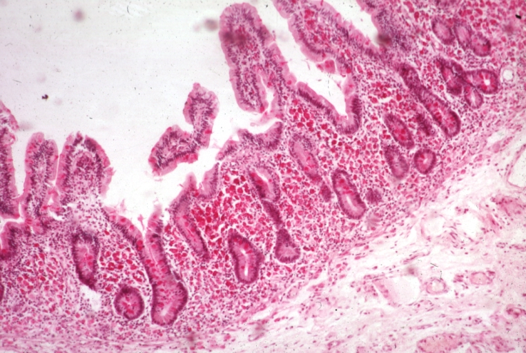
Malabsorption
Wikipedia
See also [ edit ] Fructose malabsorption Protein losing enteropathy References [ edit ] ^ a b c d e "Malabsorption Syndrome" . MedlinePlus . Retrieved 29 April 2018 . ^ Fine, KD; Schiller, LR (1999). ... PMID 10348832 . ^ a b c Bai J (1998). "Malabsorption syndromes". Digestion . 59 (5): 530–46. doi : 10.1159/000007529 . ... S2CID 46786949 . ^ health a to z "Malabsorption syndrome" . Archived from the original on 2007-05-22 . ... "Chronic diarrhea and malabsorption (including short gut syndrome): Working Group Report of the First World Congress of Pediatric Gastroenterology, Hepatology, and Nutrition". ... External links [ edit ] Classification D ICD - 10 : ( K90 ) ICD - 9-CM : 579 MeSH : D008286 DiseasesDB : 7698 External resources MedlinePlus : 000299 eMedicine : med/1384 v t e Diseases of the digestive system Upper GI tract Esophagus Esophagitis Candidal Eosinophilic Herpetiform Rupture Boerhaave syndrome Mallory–Weiss syndrome UES Zenker's diverticulum LES Barrett's esophagus Esophageal motility disorder Nutcracker esophagus Achalasia Diffuse esophageal spasm Gastroesophageal reflux disease (GERD) Laryngopharyngeal reflux (LPR) Esophageal stricture Megaesophagus Esophageal intramural pseudodiverticulosis Stomach Gastritis Atrophic Ménétrier's disease Gastroenteritis Peptic (gastric) ulcer Cushing ulcer Dieulafoy's lesion Dyspepsia Pyloric stenosis Achlorhydria Gastroparesis Gastroptosis Portal hypertensive gastropathy Gastric antral vascular ectasia Gastric dumping syndrome Gastric volvulus Buried bumper syndrome Gastrinoma Zollinger–Ellison syndrome Lower GI tract Enteropathy Small intestine ( Duodenum / Jejunum / Ileum ) Enteritis Duodenitis Jejunitis Ileitis Peptic (duodenal) ulcer Curling's ulcer Malabsorption : Coeliac Tropical sprue Blind loop syndrome Small bowel bacterial overgrowth syndrome Whipple's Short bowel syndrome Steatorrhea Milroy disease Bile acid malabsorption Large intestine ( Appendix / Colon ) Appendicitis Colitis Pseudomembranous Ulcerative Ischemic Microscopic Collagenous Lymphocytic Functional colonic disease IBS Intestinal pseudoobstruction / Ogilvie syndrome Megacolon / Toxic megacolon Diverticulitis / Diverticulosis / SCAD Large and/or small Enterocolitis Necrotizing Gastroenterocolitis IBD Crohn's disease Vascular : Abdominal angina Mesenteric ischemia Angiodysplasia Bowel obstruction : Ileus Intussusception Volvulus Fecal impaction Constipation Diarrhea Infectious Intestinal adhesions Rectum Proctitis Radiation proctitis Proctalgia fugax Rectal prolapse Anismus Anal canal Anal fissure / Anal fistula Anal abscess Hemorrhoid Anal dysplasia Pruritus ani GI bleeding Blood in stool Upper Hematemesis Melena Lower Hematochezia Accessory Liver Hepatitis Viral hepatitis Autoimmune hepatitis Alcoholic hepatitis Cirrhosis PBC Fatty liver NASH Vascular Budd–Chiari syndrome Hepatic veno-occlusive disease Portal hypertension Nutmeg liver Alcoholic liver disease Liver failure Hepatic encephalopathy Acute liver failure Liver abscess Pyogenic Amoebic Hepatorenal syndrome Peliosis hepatis Metabolic disorders Wilson's disease Hemochromatosis Gallbladder Cholecystitis Gallstone / Cholelithiasis Cholesterolosis Adenomyomatosis Postcholecystectomy syndrome Porcelain gallbladder Bile duct / Other biliary tree Cholangitis Primary sclerosing cholangitis Secondary sclerosing cholangitis Ascending Cholestasis / Mirizzi's syndrome Biliary fistula Haemobilia Common bile duct Choledocholithiasis Biliary dyskinesia Sphincter of Oddi dysfunction Pancreatic Pancreatitis Acute Chronic Hereditary Pancreatic abscess Pancreatic pseudocyst Exocrine pancreatic insufficiency Pancreatic fistula Other Hernia Diaphragmatic Congenital Hiatus Inguinal Indirect Direct Umbilical Femoral Obturator Spigelian Lumbar Petit's Grynfeltt-Lesshaft Undefined location Incisional Internal hernia Richter's Peritoneal Peritonitis Spontaneous bacterial peritonitis Hemoperitoneum PneumoperitoneumSLC46A1, CFTR, CBLIF, SI, MTTP, NEUROG3, ARX, SLC5A1, PCSK1, SLC6A19, RFX6, PEX14, PEX6, TCF3, PEX10, TERC, TERT, TGFB1, TGFBR2, TLR4, KDM6A, CLIP2, STAT4, WFS1, XRCC4, KMT2D, KLRC4, PEX3, RFXANK, PEX11B, BAZ1B, STX1A, AKR1D1, SRP54, RFC2, PIK3CA, PEX13, PIK3R1, PMS1, PMS2, POLG, PEX19, PEX5, RFX5, ABCB4, RFXAP, RMRP, RPS20, SAA1, PEX12, PEX16, SLC2A2, SLCO2A1, RECQL4, AGA, ADAMTS3, USB1, SLC39A4, NHP2, PEX26, LRRC8A, TRMT5, SEMA4A, CYBC1, EFL1, FAT4, CTC1, GTF2IRD1, HSD3B7, CDCA7, DNAJC21, CCBE1, IL23R, UBR1, UBAC2, CISD2, NCF1, NOP10, SLC29A3, WRAP53, NSUN2, LPIN2, PEX1, HYOU1, SRCAP, SPINK5, CLCA4, FAN1, PMPCA, VPS13A, TINF2, TBL2, MLH3, BLNK, SH3KBP1, FOXP3, SBDS, DCTN4, RTEL1, ERAP1, ZBTB24, PEX2, ENPP1, DNMT3B, ELN, ERCC2, F5, FLNA, GLA, MSH6, GTF2I, HBB, HELLS, HLA-B, HLA-DRB1, PARN, IGHM, IGLL1, IL10, IL12A, IRF5, JAK2, KRAS, TYMP, DMP1, LIMK1, DKC1, AK2, APC, FAS, ATP7A, B2M, BMPR1A, BTK, C4A, CAV1, CD79A, CD79B, CCR1, CCR6, CCN2, CTNNB1, CYBA, CYBB, CYP27A1, CD55, LIG4, HPGD, IL12A-AS1, TRNW, COX1, COX2, COX3, ND1, MYD88, ND5, ND6, TRNE, MPI, TRNS2, TRNF, TRNH, TRNK, TRNL1, TRNQ, TRNS1, MSH2, ND4, MLH1, MEFV, NCF4, OCRL, EPCAM, NCF2, CIITA, CUBN, SLC2A5, APOB, SAR1B, TNF, TGM2, ACE, AGT, ACAD8, AMN, PTH, GCG, MMUT, PAEP, MYO5B, HT, ATP4A, MYLK, TPH2, PLF, PYY, HAMP, NLRP3, ATP12A, COX4I2, PNLIP, OR10A4, CD40, PANK2, REXO1L1P, CD36, ATP8B1, CCK, TPH1, CLDN4, PDP1, UCP2, EZR, LEP, LCT, TH, KIT, KDR, DGAT1, RAB11A, NRP1, CLDN2, MT1B, NCOA2, SLC10A2, MGLL, GSR, GH1, PTPN22, CNNM4, FGR, FAT1, FABP1, MTR, EXOSC3, TTR, S100A10, RNPEP, MLXIPL
-
Oculogyric Crisis
Wikipedia
The abrupt termination of the psychiatric symptoms at the conclusion of the crisis is most striking. [ citation needed ] Other features that are noted during attacks include mutism , palilalia , eye blinking, lacrimation , pupil dilation , drooling, respiratory dyskinesia , increased blood pressure and heart rate, facial flushing, headache, vertigo, anxiety, agitation, compulsive thinking, paranoia, depression, recurrent fixed ideas, depersonalization, violence, and obscene language. [ citation needed ] It is often not realized that in addition to the acute presentation, oculogyric crisis can develop as a recurrent syndrome, triggered by stress, and exposure to the drugs mentioned below. [ citation needed ] Causes [ edit ] Drugs that can trigger an oculogyric crisis include neuroleptics (such as haloperidol , chlorpromazine , fluphenazine , olanzapine ), [3] carbamazepine , chloroquine , cisplatin , diazoxide , levodopa , [4] lithium , metoclopramide , lurasidone , domperidone , nifedipine , pemoline , [ citation needed ] phencyclidine ("PCP") , [5] reserpine , and cetirizine , an antihistamine. ... Other causes can include Aromatic L-amino acid decarboxylase deficiency, [6] postencephalitic Parkinson's , Tourette's syndrome , multiple sclerosis , neurosyphilis , head trauma, bilateral thalamic infarction , lesions of the fourth ventricle , cystic glioma of the third ventricle , herpes encephalitis , kernicterus and juvenile Parkinson's . ... External links [ edit ] Classification D ICD - 10 : H51.8 ICD - 9-CM : 378.87 DiseasesDB : 34992 External resources eMedicine : emerg/338 v t e Diseases of the human eye Adnexa Eyelid Inflammation Stye Chalazion Blepharitis Entropion Ectropion Lagophthalmos Blepharochalasis Ptosis Blepharophimosis Xanthelasma Ankyloblepharon Eyelash Trichiasis Madarosis Lacrimal apparatus Dacryoadenitis Epiphora Dacryocystitis Xerophthalmia Orbit Exophthalmos Enophthalmos Orbital cellulitis Orbital lymphoma Periorbital cellulitis Conjunctiva Conjunctivitis allergic Pterygium Pseudopterygium Pinguecula Subconjunctival hemorrhage Globe Fibrous tunic Sclera Scleritis Episcleritis Cornea Keratitis herpetic acanthamoebic fungal Exposure Photokeratitis Corneal ulcer Thygeson's superficial punctate keratopathy Corneal dystrophy Fuchs' Meesmann Corneal ectasia Keratoconus Pellucid marginal degeneration Keratoglobus Terrien's marginal degeneration Post-LASIK ectasia Keratoconjunctivitis sicca Corneal opacity Corneal neovascularization Kayser–Fleischer ring Haab's striae Arcus senilis Band keratopathy Vascular tunic Iris Ciliary body Uveitis Intermediate uveitis Hyphema Rubeosis iridis Persistent pupillary membrane Iridodialysis Synechia Choroid Choroideremia Choroiditis Chorioretinitis Lens Cataract Congenital cataract Childhood cataract Aphakia Ectopia lentis Retina Retinitis Chorioretinitis Cytomegalovirus retinitis Retinal detachment Retinoschisis Ocular ischemic syndrome / Central retinal vein occlusion Central retinal artery occlusion Branch retinal artery occlusion Retinopathy diabetic hypertensive Purtscher's of prematurity Bietti's crystalline dystrophy Coats' disease Sickle cell Macular degeneration Retinitis pigmentosa Retinal haemorrhage Central serous retinopathy Macular edema Epiretinal membrane (Macular pucker) Vitelliform macular dystrophy Leber's congenital amaurosis Birdshot chorioretinopathy Other Glaucoma / Ocular hypertension / Primary juvenile glaucoma Floater Leber's hereditary optic neuropathy Red eye Globe rupture Keratomycosis Phthisis bulbi Persistent fetal vasculature / Persistent hyperplastic primary vitreous Persistent tunica vasculosa lentis Familial exudative vitreoretinopathy Pathways Optic nerve Optic disc Optic neuritis optic papillitis Papilledema Foster Kennedy syndrome Optic atrophy Optic disc drusen Optic neuropathy Ischemic anterior (AION) posterior (PION) Kjer's Leber's hereditary Toxic and nutritional Strabismus Extraocular muscles Binocular vision Accommodation Paralytic strabismus Ophthalmoparesis Chronic progressive external ophthalmoplegia Kearns–Sayre syndrome palsies Oculomotor (III) Fourth-nerve (IV) Sixth-nerve (VI) Other strabismus Esotropia / Exotropia Hypertropia Heterophoria Esophoria Exophoria Cyclotropia Brown's syndrome Duane syndrome Other binocular Conjugate gaze palsy Convergence insufficiency Internuclear ophthalmoplegia One and a half syndrome Refraction Refractive error Hyperopia Myopia Astigmatism Anisometropia / Aniseikonia Presbyopia Vision disorders Blindness Amblyopia Leber's congenital amaurosis Diplopia Scotoma Color blindness Achromatopsia Dichromacy Monochromacy Nyctalopia Oguchi disease Blindness / Vision loss / Visual impairment Anopsia Hemianopsia binasal bitemporal homonymous Quadrantanopia subjective Asthenopia Hemeralopia Photophobia Scintillating scotoma Pupil Anisocoria Argyll Robertson pupil Marcus Gunn pupil Adie syndrome Miosis Mydriasis Cycloplegia Parinaud's syndrome Other Nystagmus Childhood blindness Infections Trachoma Onchocerciasis
-
Accelerated Idioventricular Rhythm
Wikipedia
However, ventricular tachycardia and ventricular fibrillation remain the most important causes of sudden death following spontaneous restoration of antegrade flow. [6] Prior to the modern practice of percutaneous coronary intervention for acute coronary syndrome , pharmacologic thrombolysis was more common and accelerated idioventricular rhythms were used as a sign of successful reperfusion. [7] It is considered a benign arrhythmia especially in the setting of STEMI(where it is conventionally thought to be an indicator of reperfusion) that does not require intervention, though atrioventricular dyssynchrony can cause hemodynamic instability, which can be treated through overdrive pacing or atropine . [2] Diagnosis [ edit ] Differential diagnosis [ edit ] AIVR appears similar to ventricular tachycardia with wide QRS complexes (QRS >0.12s) and a regular rhythm. ... CS1 maint: extra text: authors list ( link ) v t e Cardiovascular disease (heart) Ischaemic Coronary disease Coronary artery disease (CAD) Coronary artery aneurysm Spontaneous coronary artery dissection (SCAD) Coronary thrombosis Coronary vasospasm Myocardial bridge Active ischemia Angina pectoris Prinzmetal's angina Stable angina Acute coronary syndrome Myocardial infarction Unstable angina Sequelae hours Hibernating myocardium Myocardial stunning days Myocardial rupture weeks Aneurysm of heart / Ventricular aneurysm Dressler syndrome Layers Pericardium Pericarditis Acute Chronic / Constrictive Pericardial effusion Cardiac tamponade Hemopericardium Myocardium Myocarditis Chagas disease Cardiomyopathy Dilated Alcoholic Hypertrophic Tachycardia-induced Restrictive Loeffler endocarditis Cardiac amyloidosis Endocardial fibroelastosis Arrhythmogenic right ventricular dysplasia Endocardium / valves Endocarditis infective endocarditis Subacute bacterial endocarditis non-infective endocarditis Libman–Sacks endocarditis Nonbacterial thrombotic endocarditis Valves mitral regurgitation prolapse stenosis aortic stenosis insufficiency tricuspid stenosis insufficiency pulmonary stenosis insufficiency Conduction / arrhythmia Bradycardia Sinus bradycardia Sick sinus syndrome Heart block : Sinoatrial AV 1° 2° 3° Intraventricular Bundle branch block Right Left Left anterior fascicle Left posterior fascicle Bifascicular Trifascicular Adams–Stokes syndrome Tachycardia ( paroxysmal and sinus ) Supraventricular Atrial Multifocal Junctional AV nodal reentrant Junctional ectopic Ventricular Accelerated idioventricular rhythm Catecholaminergic polymorphic Torsades de pointes Premature contraction Atrial Junctional Ventricular Pre-excitation syndrome Lown–Ganong–Levine Wolff–Parkinson–White Flutter / fibrillation Atrial flutter Ventricular flutter Atrial fibrillation Familial Ventricular fibrillation Pacemaker Ectopic pacemaker / Ectopic beat Multifocal atrial tachycardia Pacemaker syndrome Parasystole Wandering atrial pacemaker Long QT syndrome Andersen–Tawil Jervell and Lange-Nielsen Romano–Ward Cardiac arrest Sudden cardiac death Asystole Pulseless electrical activity Sinoatrial arrest Other / ungrouped hexaxial reference system Right axis deviation Left axis deviation QT Short QT syndrome T T wave alternans ST Osborn wave ST elevation ST depression Strain pattern Cardiomegaly Ventricular hypertrophy Left Right / Cor pulmonale Atrial enlargement Left Right Athletic heart syndrome Other Cardiac fibrosis Heart failure Diastolic heart failure Cardiac asthma Rheumatic fever External links [ edit ] Classification D ICD - 10 : I45.6 MeSH : D016170 DiseasesDB : 31195

-
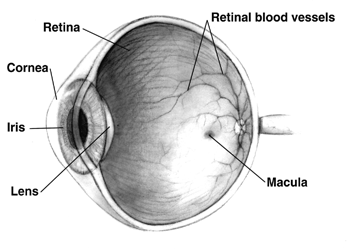
Purtscher's Retinopathy
Wikipedia
Contents 1 Presentation 1.1 Associated diseases 2 Pathophysiology 3 Diagnosis 4 Treatment 5 Prognosis 6 History 7 References 8 External links Presentation [ edit ] Associated diseases [ edit ] Severe head , chest , or long bone trauma Acute pancreatitis Amniotic fluid embolism Chronic kidney failure Dermatomyositis Fat embolism syndrome Scleroderma Systemic lupus erythematosus (SLE) Thrombotic thrombocytopenic purpura (TTP) Pathophysiology [ edit ] Purtscher's retinopathy likely involves complex pathophysiology, with several contributing factors, including complement -mediated aggregates, fat, air, fibrin clots and platelet clumps. [1] The diseases leads to the formation of cotton wool spots in the retina, a finding observed in several other diseases, and atrophy of the optic nerve . [2] Diagnosis [ edit ] Where trauma is involved, only a funduscopic examination of the back of the eye (retina) is necessary to make the diagnosis. [2] Fluoroscein angiography may show a decrease in blood flow to the areas of whiteness in the retina. ... Prognosis [ edit ] Purtscher's retinopathy can lead to loss of vision, [4] and recovery of vision may occur very little. [2] However, vision recovery does occur in some cases, and reports have varied on the long-term prognosis. [5] History [ edit ] Purtscher's retinopathy was first characterized in 1910 and 1912 as a syndrome of sudden blindness after head trauma , [6] with patches of hemorrhage and whitening of the retina in both eyes. [2] Later, it was discovered to occur after other types of trauma , such as chest trauma, and is associated with several non-traumatic systemic diseases. [2] Purtscher's retinopathy may also be associated with acute pancreatitis , vasculitis , embolization of such materials as fat and amniotic fluid , [7] systemic lupus erythematosus, thrombotic thrombocytopenic purpura, and chronic kidney failure. [2] Purtscher's retinopathy may be caused by extensive fractures of the long bones. [8] References [ edit ] ^ Buckley, Sally A.; James, B (July 1996). ... External links [ edit ] Classification D DiseasesDB : 33929 External resources eMedicine : article/1225431 v t e Diseases of the human eye Adnexa Eyelid Inflammation Stye Chalazion Blepharitis Entropion Ectropion Lagophthalmos Blepharochalasis Ptosis Blepharophimosis Xanthelasma Ankyloblepharon Eyelash Trichiasis Madarosis Lacrimal apparatus Dacryoadenitis Epiphora Dacryocystitis Xerophthalmia Orbit Exophthalmos Enophthalmos Orbital cellulitis Orbital lymphoma Periorbital cellulitis Conjunctiva Conjunctivitis allergic Pterygium Pseudopterygium Pinguecula Subconjunctival hemorrhage Globe Fibrous tunic Sclera Scleritis Episcleritis Cornea Keratitis herpetic acanthamoebic fungal Exposure Photokeratitis Corneal ulcer Thygeson's superficial punctate keratopathy Corneal dystrophy Fuchs' Meesmann Corneal ectasia Keratoconus Pellucid marginal degeneration Keratoglobus Terrien's marginal degeneration Post-LASIK ectasia Keratoconjunctivitis sicca Corneal opacity Corneal neovascularization Kayser–Fleischer ring Haab's striae Arcus senilis Band keratopathy Vascular tunic Iris Ciliary body Uveitis Intermediate uveitis Hyphema Rubeosis iridis Persistent pupillary membrane Iridodialysis Synechia Choroid Choroideremia Choroiditis Chorioretinitis Lens Cataract Congenital cataract Childhood cataract Aphakia Ectopia lentis Retina Retinitis Chorioretinitis Cytomegalovirus retinitis Retinal detachment Retinoschisis Ocular ischemic syndrome / Central retinal vein occlusion Central retinal artery occlusion Branch retinal artery occlusion Retinopathy diabetic hypertensive Purtscher's of prematurity Bietti's crystalline dystrophy Coats' disease Sickle cell Macular degeneration Retinitis pigmentosa Retinal haemorrhage Central serous retinopathy Macular edema Epiretinal membrane (Macular pucker) Vitelliform macular dystrophy Leber's congenital amaurosis Birdshot chorioretinopathy Other Glaucoma / Ocular hypertension / Primary juvenile glaucoma Floater Leber's hereditary optic neuropathy Red eye Globe rupture Keratomycosis Phthisis bulbi Persistent fetal vasculature / Persistent hyperplastic primary vitreous Persistent tunica vasculosa lentis Familial exudative vitreoretinopathy Pathways Optic nerve Optic disc Optic neuritis optic papillitis Papilledema Foster Kennedy syndrome Optic atrophy Optic disc drusen Optic neuropathy Ischemic anterior (AION) posterior (PION) Kjer's Leber's hereditary Toxic and nutritional Strabismus Extraocular muscles Binocular vision Accommodation Paralytic strabismus Ophthalmoparesis Chronic progressive external ophthalmoplegia Kearns–Sayre syndrome palsies Oculomotor (III) Fourth-nerve (IV) Sixth-nerve (VI) Other strabismus Esotropia / Exotropia Hypertropia Heterophoria Esophoria Exophoria Cyclotropia Brown's syndrome Duane syndrome Other binocular Conjugate gaze palsy Convergence insufficiency Internuclear ophthalmoplegia One and a half syndrome Refraction Refractive error Hyperopia Myopia Astigmatism Anisometropia / Aniseikonia Presbyopia Vision disorders Blindness Amblyopia Leber's congenital amaurosis Diplopia Scotoma Color blindness Achromatopsia Dichromacy Monochromacy Nyctalopia Oguchi disease Blindness / Vision loss / Visual impairment Anopsia Hemianopsia binasal bitemporal homonymous Quadrantanopia subjective Asthenopia Hemeralopia Photophobia Scintillating scotoma Pupil Anisocoria Argyll Robertson pupil Marcus Gunn pupil Adie syndrome Miosis Mydriasis Cycloplegia Parinaud's syndrome Other Nystagmus Childhood blindness Infections Trachoma Onchocerciasis
-
Trochleitis
Wikipedia
Inflammation of the trochlear region leads to a painful syndrome with swelling and exquisite point tenderness in the upper medial rim of the orbit. ... History [ edit ] Trochleitis was first identified in 1984 by Tychsen, et al. in a study of thirteen patients with orbital pain and point tenderness over the trochlear region. Previously, the trochleitis syndrome had been included in the broad category of idiopathic orbital inflammation (also called orbital pseudotumor). ... Neurology 2004: 62:1134-1140. v t e Diseases of the human eye Adnexa Eyelid Inflammation Stye Chalazion Blepharitis Entropion Ectropion Lagophthalmos Blepharochalasis Ptosis Blepharophimosis Xanthelasma Ankyloblepharon Eyelash Trichiasis Madarosis Lacrimal apparatus Dacryoadenitis Epiphora Dacryocystitis Xerophthalmia Orbit Exophthalmos Enophthalmos Orbital cellulitis Orbital lymphoma Periorbital cellulitis Conjunctiva Conjunctivitis allergic Pterygium Pseudopterygium Pinguecula Subconjunctival hemorrhage Globe Fibrous tunic Sclera Scleritis Episcleritis Cornea Keratitis herpetic acanthamoebic fungal Exposure Photokeratitis Corneal ulcer Thygeson's superficial punctate keratopathy Corneal dystrophy Fuchs' Meesmann Corneal ectasia Keratoconus Pellucid marginal degeneration Keratoglobus Terrien's marginal degeneration Post-LASIK ectasia Keratoconjunctivitis sicca Corneal opacity Corneal neovascularization Kayser–Fleischer ring Haab's striae Arcus senilis Band keratopathy Vascular tunic Iris Ciliary body Uveitis Intermediate uveitis Hyphema Rubeosis iridis Persistent pupillary membrane Iridodialysis Synechia Choroid Choroideremia Choroiditis Chorioretinitis Lens Cataract Congenital cataract Childhood cataract Aphakia Ectopia lentis Retina Retinitis Chorioretinitis Cytomegalovirus retinitis Retinal detachment Retinoschisis Ocular ischemic syndrome / Central retinal vein occlusion Central retinal artery occlusion Branch retinal artery occlusion Retinopathy diabetic hypertensive Purtscher's of prematurity Bietti's crystalline dystrophy Coats' disease Sickle cell Macular degeneration Retinitis pigmentosa Retinal haemorrhage Central serous retinopathy Macular edema Epiretinal membrane (Macular pucker) Vitelliform macular dystrophy Leber's congenital amaurosis Birdshot chorioretinopathy Other Glaucoma / Ocular hypertension / Primary juvenile glaucoma Floater Leber's hereditary optic neuropathy Red eye Globe rupture Keratomycosis Phthisis bulbi Persistent fetal vasculature / Persistent hyperplastic primary vitreous Persistent tunica vasculosa lentis Familial exudative vitreoretinopathy Pathways Optic nerve Optic disc Optic neuritis optic papillitis Papilledema Foster Kennedy syndrome Optic atrophy Optic disc drusen Optic neuropathy Ischemic anterior (AION) posterior (PION) Kjer's Leber's hereditary Toxic and nutritional Strabismus Extraocular muscles Binocular vision Accommodation Paralytic strabismus Ophthalmoparesis Chronic progressive external ophthalmoplegia Kearns–Sayre syndrome palsies Oculomotor (III) Fourth-nerve (IV) Sixth-nerve (VI) Other strabismus Esotropia / Exotropia Hypertropia Heterophoria Esophoria Exophoria Cyclotropia Brown's syndrome Duane syndrome Other binocular Conjugate gaze palsy Convergence insufficiency Internuclear ophthalmoplegia One and a half syndrome Refraction Refractive error Hyperopia Myopia Astigmatism Anisometropia / Aniseikonia Presbyopia Vision disorders Blindness Amblyopia Leber's congenital amaurosis Diplopia Scotoma Color blindness Achromatopsia Dichromacy Monochromacy Nyctalopia Oguchi disease Blindness / Vision loss / Visual impairment Anopsia Hemianopsia binasal bitemporal homonymous Quadrantanopia subjective Asthenopia Hemeralopia Photophobia Scintillating scotoma Pupil Anisocoria Argyll Robertson pupil Marcus Gunn pupil Adie syndrome Miosis Mydriasis Cycloplegia Parinaud's syndrome Other Nystagmus Childhood blindness Infections Trachoma Onchocerciasis
-
Northern Epilepsy Syndrome
Wikipedia
Northern epilepsy syndrome Other names Neuronal ceroid lipofuscinosis, Northern epilepsy variant This condition is inherited in an autosomal recessive manner. Northern epilepsy syndrome ( NE ), or progressive epilepsy with mental retardation ( EPMR ), is a subtype of neuronal ceroid lipofuscinosis and a rare disease that is regarded as a Finnish heritage disease . Unlike most Finnish heritage diseases, this syndrome has been reported only in Finland . [1] The disease is characterized by seizures in early childhood that progressively get worse until after puberty. ... Life expectancy is at least 50 years of age, which is shorter than the average worldwide age of 70. [4] Genetic causes [ edit ] Northern epilepsy syndrome is caused by an inherited autosomal recessive mutation in the telomeric region of the short arm of chromosome 8 . ... The disease is now known as the mildest form of NCL. [7] There are two forms of this mutated gene: 1-CLN8 and 2-CLN8. 1-CLN8 is known as Northern epilepsy syndrome, while 2-CLN8 is primarily from Turkish descent. [2] See also [ edit ] Epilepsy Neuronal ceroid lipofuscinosis CLN8 References [ edit ] ^ Krystyna E.

-
Microcephaly, Macrotia, And Mental Retardation
Omim
The authors proposed that this complex may be a previously undescribed syndrome with autosomal dominant or X-linked dominant inheritance. The mild form of Cornelia de Lange syndrome (122470) and the Kabuki syndrome (147920) have some overlapping facial features with this entity.
-
Reunion Island Larsen-Like Syndrome
Orphanet
A rare, genetic, congenital disorder of glycosylation characterized by severe, pre- and post-natal short stature, joint hyperlaxity with multiple dislocations (elbows, fingers, hips, knees), and facial dysmorphism (round flat face, high forehead, hypertelorism, prominent bulging eyes with under-eye shadows, hypoplastic midface, microstomia, protruding lips). Other associated features may include cutaneous hyperextensibility, learning difficulties, and ocular abnormalities. Advanced carpal ossification, widened metaphyses, and, occasionally, radioulnar synostosis, scoliosis and a Swedish key appearance of the proximal femora, is observed on imaging.
-
Aneurysm-Osteoarthritis Syndrome
Orphanet
A rare, genetic, systemic disease characterized by the presence of arterial aneurysms, tortuosity and dissection throughout the arterial tree, associated with early-onset osteoarthritis (predominantly affecting the spine, hands and/or wrists, and knees) and mild craniofacial dysmorphism (incl. long face, high forehead, flat supraorbital ridges, hypertelorism, malar hypoplasia and, a raphe, broad or bifid uvula), as well as mild skeletal and cutaneous anomalies. Joint abnormalities, such as osteochondritis dissecans and intervertebral disc degeneration, are frequently associated. Additonal cardiovascular anomalies may include mitral valve defects, congenital heart malformations, ventricular hypertrophy and atrial fibrillation.
-
Autosomal Dominant Myopia-Midfacial Retrusion-Sensorineural Hearing Loss-Rhizomelic Dysplasia Syndrome
Orphanet
A rare primary bone dysplasia characterized by micromelia with rhizomelic shortening, metaphyseal widening of the long bones, brachydactyly, small scapulae, micrognathia and thoracic insufficiency requiring tracheostomy and ventilation, and severe myopia and sensorineural hearing loss. Further dysmorphic craniofacial features include frontal bossing, proptosis, epicanthal folds, short nose, flat nasal bridge, anteverted nares, midfacial retrusion, and cleft palate.
-
Autosomal Dominant Distal Axonal Motor Neuropathy-Myofibrillar Myopathy Syndrome
Orphanet
A rare genetic neuromuscular disease characterized by length-dependent axonal motor neuropathy predominantly affecting the lower limbs, in combination with a myopathy with morphological features of myofibrillar myopathy with aggregates and rimmed vacuoles. Age of onset is typically in the second to third decade of life. Patients present with slowly progressive muscle weakness and atrophy initially affecting the distal lower limbs and later progressing to involve proximal limbs and also truncal muscles. There is no involvement of respiratory and cardiac muscles.
-
Autosomal Recessive Cerebellar Ataxia-Pyramidal Signs-Nystagmus-Oculomotor Apraxia Syndrome
Orphanet
A rare, genetic, slowly progressive neurodegenerative disease characterized by delayed psychomotor development beginning in infancy, mild to profound intellectual disability, gait and stance ataxia, pyramidal signs (hyperreflexia, extensor plantar responses), dysarthria, and ocular abnormalities (e.g. nystagmus, oculomotor apraxia, abduction deficits, esotropia, ptosis). Brain imaging reveals progressive, generalized cerebellar atrophy, mild ventriculomegaly and, in some, retrocerebellar cysts.
-
Optic Atrophy-Ataxia-Peripheral Neuropathy-Global Developmental Delay Syndrome
Orphanet
A rare mitochondrial disease characterized by a variable clinical phenotype with the core features of optic atrophy, ataxia, and hypotonia. Additional common manifestations include global developmental delay with or without regression, neuropathy, spasticity, and microcephaly, less frequently seizures, movement disorder, hearing loss, and respiratory failure. Brain imaging may show abnormalities of the corpus callosum, basal ganglia, and midbrain, cerebral or cerebellar atrophy, or white matter abnormalities. The condition is frequently fatal at an early age.
-
Neonatal Diabetes-Congenital Hypothyroidism-Congenital Glaucoma-Hepatic Fibrosis-Polycystic Kidneys Syndrome
Orphanet
A rare genetic disease characterized by intrauterine growth retardation, permanent neonatal diabetes mellitus, and congenital hypothyroidism. Additional manifestations include congenital glaucoma, hepatic disease (hepatitis, fibrosis, and cirrhosis), polycystic kidneys, exocrine pancreatic dysfunction, sensorineural hearing impairment, developmental delay, and mild facial dysmorphism (such as flat nasal bridge, epicanthal folds, long philtrum, and low-set ears), among others.
-
Pterygium Colli-Intellectual Disability-Digital Anomalies Syndrome
Orphanet
A rare disorder characterized by pterygium colli, digital anomalies (abnormal small thumbs, widened interphalangeal joints, and broad terminal phalanges), and craniofacial abnormalities (brachycephaly, epicanthic folds, angulated eyebrows, upward slanting of the palpebral fissures, ptosis, hypertelorism, and prominent low-set, posteriorly rotated ears). It has been described in a woman and her son, but the manifestations were much less severe in the mother. The son also had intellectual deficit. The inheritance is either X-linked dominant or autosomal dominant.
-
Autism Spectrum Disorder-Epilepsy-Arthrogryposis Syndrome
Orphanet
SLC35A3-CDG is a form of congenital disorders of N-linked glycosylation characterized by distal arthrogryposis (mild flexion contractures of the fingers, deviation of the distal phalanges, swan-neck deformity), retromicrognathia, general muscle hypotonia, delayed psychomotor development, autism spectrum disorder (speech delay, abnormal use of speech, difficulties in initiating, understanding and maintaining social interaction, limited non-verbal communication and repetitive behavior), seizures, microcephaly and mild to moderate intellectual disability that becomes apparent with age. The disease is caused by mutations in the gene SLC35A3 (1p21).
-
Glossopalatine Ankylosis
Orphanet
Glossopalatine ankylosis is a disorder belonging to the group of oromandibular-limb hypogenesis syndromes (OLHS) and is characterised by the presence of an intraoral band of variable thickness attaching the tongue to the hard palate or maxillary alveolar ridge. Epidemiology The syndrome is very rare with less than 30 cases reported in the literature so far. ... Etiology The aetiology is unknown and the syndrome appears to be sporadic.


